Appendix
Revision as of 10:53, 21 January 2020 by Mikael Häggström (talk | contribs) (→Microscopic evaluation: +Image)
Author:
Mikael Häggström [note 1]
Contents
Fixation
Generally 10% neutral buffered formalin.
See also: General notes on fixation
Gross processing
See also: General notes on gross processing
Microscopic evaluation

- Look for cancerous cells (also for specimens with clinical appendicitis).

Low-grade appendiceal mucinous neoplasm: Minimal cytological atypia of the epithelial cells.[1]
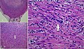
Histopathology of an appendiceal carcinoid. The arrow points out a cluster of neuroendocrine cells. There are also inflammatory cells consistent with acute appendicitis.[2]


Report
- Description of objective findings.
- Presence or absence of malignancy.
See also: General notes on reporting
Notes
- ↑ For a full list of contributors, see article history. Creators of images are attributed at the image description pages, seen by clicking on the images. See Patholines:Authorship for details.
Main page
References
- ↑ Hajjar, Roy; Dubé, Pierre; Mitchell, Andrew; Sidéris, Lucas (2019). "Combined Mucinous and Neuroendocrine Tumours of the Appendix Managed with Surgical Cytoreduction and Oxaliplatin-based Hyperthermic Intraperitoneal Chemotherapy ". Cureus. doi:. ISSN 2168-8184.
- ↑ Elkbuli, Adel; Sanchez, Carol; McKenney, Mark; Boneva, Dessy (2019). "Incidental neuro-endocrine tumor of the appendix: Case report and literature review ". Annals of Medicine and Surgery 43: 44–47. doi:. ISSN 20490801.
Image sources