Difference between revisions of "Hepatocellular carcinoma"
Jump to navigation
Jump to search
m (→Differentiation: Moved) |
(→Microscopic evaluation: +Steatotic) |
||
| Line 15: | Line 15: | ||
*Abundant granular '''eosinophilic cytoplasm''' | *Abundant granular '''eosinophilic cytoplasm''' | ||
*'''Nuclei''' with increased N/C ratio, round nuclei with coarse chromatin and thickened nuclear membrane, and may have prominent nucleoli. | *'''Nuclei''' with increased N/C ratio, round nuclei with coarse chromatin and thickened nuclear membrane, and may have prominent nucleoli. | ||
| + | *'''Steatotic''' hepatocellular carcinoma is a common variant, found most commonly in small, well-differentiated tumors.<ref name="ChanYu2016">{{cite journal|last1=Chan|first1=Anthony W H|last2=Yu|first2=Shuangni|last3=Yu|first3=Yau-Hei|last4=Tong|first4=Joanna H M|last5=Wang|first5=Lei|last6=Tin|first6=Edith K Y|last7=Chong|first7=Charing C N|last8=Chan|first8=Stephen L|last9=Wong|first9=Grace L H|last10=Wong|first10=Vincent W S|last11=Chan|first11=Henry L Y|last12=Lai|first12=Paul B S|last13=To|first13=Ka-Fai|title=Steatotic hepatocellular carcinoma: a variant associated with metabolic factors and late tumour relapse|journal=Histopathology|volume=69|issue=6|year=2016|pages=971–984|issn=03090167|doi=10.1111/his.13029}}</ref> | ||
<gallery mode=packed heights=200> | <gallery mode=packed heights=200> | ||
Revision as of 10:09, 11 February 2021
Author:
Mikael Häggström [note 1]
Contents
Presentations
Hepatocellular carcinoma is the most common diagnosis for liver tumors.:[1]
Microscopic evaluation
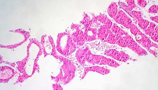
Hepatocellular carcinoma typically displays:[2]
- Trabecular pattern surrounded by layer of flattened endothelial cells.
- Presence of sinusoidal vessels surrounding tumor cells
- Scant stroma, and minimal desmoplasia
- Polygonal cells with distinct cell membranes
- Abundant granular eosinophilic cytoplasm
- Nuclei with increased N/C ratio, round nuclei with coarse chromatin and thickened nuclear membrane, and may have prominent nucleoli.
- Steatotic hepatocellular carcinoma is a common variant, found most commonly in small, well-differentiated tumors.[3]
Needle biopsy.

Needle biopsy.

Needle biopsy.

FNA.

FNA clot section.

FNA clot section.
.jpg)
.jpg)
.jpg)
.jpg)
.jpg)
.jpg)
Differentiation
Differentiations of hepatocellular carcinoma are:[2]

- Well differentiated:
- Thin plates (1 - 3 hepatocytes thick)
- Hepatocytes are smaller than normal
- Abnormal reticulin network
- Minimal nuclear atypia
- Nuclear density of 2x compared to normal liver

- Moderately differentiated:
- Trabecular pattern at least 4 cells thick
- Larger cells with more eosinophilic cytoplasm
- Distinct nucleoli
- Pseudoglands

- Poorly differentiated:
- Large tumor cells with hyperchromatic nuclei in compact growth pattern
- Rare trabeculae or bile
- Prominent pleomorphism
- May have spindle cell or small cell areas
Notes
- ↑ For a full list of contributors, see article history. Creators of images are attributed at the image description pages, seen by clicking on the images. See Patholines:Authorship for details.
Main page
References
- ↑ Table 37.2 in: Sternberg, Stephen (2012). Sternberg's diagnostic surgical pathology . Place of publication not identified: LWW. ISBN 978-1-4511-5289-0. OCLC 953861627.
- ↑ 2.0 2.1 Deepali Jain. Liver & intrahepatic bile ducts - Hepatocellular carcinoma - Hepatocellular carcinoma overview. PathologyOutlines. Topic Completed: 1 February 2012. Minor changes: 30 September 2020
- ↑ Chan, Anthony W H; Yu, Shuangni; Yu, Yau-Hei; Tong, Joanna H M; Wang, Lei; Tin, Edith K Y; Chong, Charing C N; Chan, Stephen L; et al. (2016). "Steatotic hepatocellular carcinoma: a variant associated with metabolic factors and late tumour relapse ". Histopathology 69 (6): 971–984. doi:. ISSN 03090167.
Image sources