Ovary
Jump to navigation
Jump to search

Author:
Mikael Häggström [note 1]
Contents
Fixation
Generally 10% neutral buffered formalin.
Presentations
Gross processing

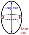
"Long" and "short" axis.[1]
The ovary is cut in the longitudinal plane (through the "long axis").
Ovaries, including those with cysts, are almost never inked.
Gross report
Template:
| (A. Labeled - __. The specimen is received in formalin and consists of) an ovary measuring ___. The ovarian capsule is tan-pink and smooth. Cut sections reveal solid, white and whorled parenchyma, and no gross lesions. Representative sections are submitted for microscopic examination in __ cassettes. |
Microscopic examination

Signet ring cell carcinoma metastasis to the ovary, also called Krukenberg tumor: Gross pathology (top, cross-section at right) and histopathology at low and high magnification.[2]
Apart from any obvious tumor, also look for signet ring cells, which is a major feature of metastatic tumors to the ovary.
See also
Notes
- ↑ For a full list of contributors, see article history. Creators of images are attributed at the image description pages, seen by clicking on the images. See Patholines:Authorship for details.
Main page
References
- ↑ Pellerito, John; Polak, Joseph F. (2012). Introduction to Vascular Ultrasonography (6th ed.). Elsevier Health Sciences. p. 559. ISBN 978-1-4557-3766-6.
- ↑ Nakamura, Yoshiaki; Hiramatsu, Ayako; Koyama, Takafumi; Oyama, Yu; Tanaka, Ayuko; Honma, Koichi (2014). "A Krukenberg Tumor from an Occult Intramucosal Gastric Carcinoma Identified during an Autopsy
". Case Reports in Oncological Medicine 2014: 1–5. doi:. ISSN 2090-6706.
- Creative Commons Attribution 3.0 Unported (CC BY 3.0) license
Image sources