File:Centrocyte, centroblast and follicular dendritic cell in a follicular lymphoma.jpg
Jump to navigation
Jump to search
No higher resolution available.
Centrocyte,_centroblast_and_follicular_dendritic_cell_in_a_follicular_lymphoma.jpg (427 × 301 pixels, file size: 52 KB, MIME type: image/jpeg)
Summary
| Description |
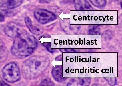
English: Histology of cell types in a germinal center: A centrocyte, a centroblast and a follicular dendritic cell, seen in a follicular lymphoma, H&E stain. - Centrocytes are small to medium size with angulated, elongated, cleaved, or twisted nuclei. - Centroblasts are larger cells containing vesicular nuclei with one to three basophilic nucleoli apposing the nuclear membrane. - Follicular dendritic cells have round nuclei, centrally located nucleoli, bland and dispersed chromatin, and flattening of adjacent nuclear membrane. Reference: Dr. William Kern. Case No.: H-003. Diagnosis: Non-Hodgkin lymphoma, follicular type, low-grade. Organ: Lymph node, inguinal. University of Oklahoma Health Sciences Center, Oklahoma. Last Updated: 12/21/2010 |
| Date | |
| Source | Own work |
| Author |
.jpg) - Reusing images - Conflicts of interest: None Consent note: Consent from the patient or patient's relatives is regarded as redundant, because of absence of identifiable features (List of HIPAA identifiers) in the media and case information (See also HIPAA case reports guidance). |
Licensing
| This file is made available under the Creative Commons CC0 1.0 Universal Public Domain Dedication. | |
| The person who associated a work with this deed has dedicated the work to the public domain by waiving all of their rights to the work worldwide under copyright law, including all related and neighboring rights, to the extent allowed by law. You can copy, modify, distribute and perform the work, even for commercial purposes, all without asking permission.
|
File history
Click on a date/time to view the file as it appeared at that time.
| Date/Time | Thumbnail | Dimensions | User | Comment | |
|---|---|---|---|---|---|
| current | 19:23, 10 November 2021 | | 427 × 301 (52 KB) | Mikael Häggström | Uploaded a work by {{Mikael Häggström|cat=Micrographs|consent=noid}} from {{Own}} with UploadWizard |
File usage
The following page uses this file: