File:Histopathology of angiolipoma.jpg
Jump to navigation
Jump to search


Size of this preview: 800 × 591 pixels. Other resolutions: 320 × 236 pixels | 640 × 473 pixels | 1,024 × 756 pixels | 1,280 × 945 pixels | 2,080 × 1,536 pixels.
Original file (2,080 × 1,536 pixels, file size: 778 KB, MIME type: image/jpeg)
Summary
| Description |
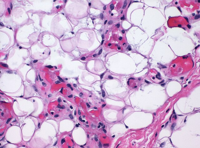
English: Histopathology of angiolipoma, high magnification, showing thick-walled capillaries with fibrin thrombi, among mature lipocytes and fibrin septa. HE stain. |
| Date | |
| Source | Own work |
| Author |
.jpg) - Reusing images - Conflicts of interest: None Consent note: Consent from the patient or patient's relatives is regarded as redundant, because of absence of identifiable features (List of HIPAA identifiers) in the media and case information (See also HIPAA case reports guidance). |
| Other versions |
 |
Licensing
| This file is made available under the Creative Commons CC0 1.0 Universal Public Domain Dedication. | |
| The person who associated a work with this deed has dedicated the work to the public domain by waiving all of their rights to the work worldwide under copyright law, including all related and neighboring rights, to the extent allowed by law. You can copy, modify, distribute and perform the work, even for commercial purposes, all without asking permission.
|
File history
Click on a date/time to view the file as it appeared at that time.
| Date/Time | Thumbnail | Dimensions | User | Comment | |
|---|---|---|---|---|---|
| current | 16:39, 9 September 2020 | | 2,080 × 1,536 (778 KB) | Mikael Häggström | Uploaded a work by {{Mikael Häggström|cat=Micrographs|consent=noid}} from {{Own}} with UploadWizard |
File usage
The following 4 pages uses this file: