Adrenals
Jump to navigation
Jump to search
Author:
Mikael Häggström [note 1]
Contents
Comprehensiveness
On this resource, the following formatting is used for comprehensiveness:
- Minimal depth
- (Moderate depth)
- ((Comprehensive))
Main targets
Autopsy
Autopsy processing
In autopsy:
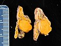
- Make a couple of cuts through the adrenal glands, such as transversal ones, and look mainly for adrenal tumors.
- ((Remove the adrenals, trim them from excessive adherent fat, and weight them. Their combined weight in an adult human ranges from 7 to 10 grams.[1]))

Adrenal cortical necrosis. Hemorrhage, fibrin thrombi and short postmortem interval indicate ante-mortem necrosis, otherwise it can be regarded as a postmortem change.[2]

Adrenal venous congestion in circulatory failure.


Autopsy report
Normal status can be described as either:
- Adrenal glands are normal bilaterally.
- (Adrenal glands are ordinarily configured and with no definable focal changes on cut surfaces.)
- ((The adrenals are normal in size, shape and consistency, with a weight of __ grams on the right and __ grams on the left. The cortices are orange with <normal / increased / decreased thickness>. The medullae are <grey / autolyzed>.))
Notes
- ↑ For a full list of contributors, see article history. Creators of images are attributed at the image description pages, seen by clicking on the images. See Patholines:Authorship for details.
Main page
References
- ↑ O'Hare, A. Munro Neville, Michael J. (1982). The Human Adrenal Cortex Pathology and Biology – An Integrated Approach . Springer London. pp. Chapter 4: Structure of the adult cortex. ISBN 9781447113171.
- ↑ Page 120 in: Rutty, Guy (2001). Essentials of autopsy practice . London New York: Springer. ISBN 978-1-85233-541-0. OCLC 44769560.
Image sources