Difference between revisions of "Appendicitis"
m (→Microscopic evaluation: mod) |
m (<noinclude>) |
||
| (33 intermediate revisions by the same user not shown) | |||
| Line 1: | Line 1: | ||
| − | {{Top | + | <noinclude>{{Top |
|author1=[[User:Mikael Häggström|Mikael Häggström]] | |author1=[[User:Mikael Häggström|Mikael Häggström]] | ||
|author2= | |author2= | ||
| − | }} | + | }}<noinclude/> |
Appendicitis may histopathologically be defined as neutrophilic infiltrates of the wall of the appendix in the correct clinical context. | Appendicitis may histopathologically be defined as neutrophilic infiltrates of the wall of the appendix in the correct clinical context. | ||
| − | |||
{{Fixation - general notes}} | {{Fixation - general notes}} | ||
| − | + | <noinclude>{{Comprehensiveness}}</noinclude> | |
| − | {{Comprehensiveness}} | ||
| − | ==Gross | + | ==Gross processing== |
| − | + | Standard sections if the appendix appears inflamed and there are no signs of malignancy. Describe abnormal signs including: | |
| − | + | <gallery mode=packed> | |
| + | File:Gross pathology of congested appendicitis.jpg|thumb|Appendicitis with '''congestion''' | ||
| + | File:Gross pathology of appendicitis with a patchy purulent exudate, annotated.jpg|Serosa with a '''patchy purulent exudate'''. | ||
| + | File:Gross pathology of appendicitis containing a blood-tinged purulent exudate.jpg|Appendicitis, with the lumen containing a '''blood-tinged purulent exudate'''. | ||
| + | File:Acute Appendicitis.jpg|Longitudinal section showing a '''red inflamed mucosa''' with an '''irregular luminal surface''' (in a case of acute suppurative appendicitis). | ||
| + | </gallery> | ||
{{Further|Appendix}} | {{Further|Appendix}} | ||
| Line 17: | Line 20: | ||
*Evaluate '''depth''' of the inflammation. | *Evaluate '''depth''' of the inflammation. | ||
*Look for any '''perforation''' of the wall. | *Look for any '''perforation''' of the wall. | ||
| + | *Look for '''cancerous cells''' (which may have caused the appendicitis). {{further|Appendix|linebreak=no}} | ||
*{{Moderate-begin}}Attempt to specify the type of appendicitis as either of the following:{{Moderate-end}} | *{{Moderate-begin}}Attempt to specify the type of appendicitis as either of the following:{{Moderate-end}} | ||
===Types=== | ===Types=== | ||
{|class=wikitable | {|class=wikitable | ||
| − | |+ Classification of acute appendicitis based on | + | |+ Classification of acute appendicitis based on gross pathology and light microscopy characteristics<ref name="Carr2000">Unless otherwise specified in rows, reference is:<br>- {{cite journal|last1=Carr|first1=Norman J.|title=The pathology of acute appendicitis|journal=Annals of Diagnostic Pathology|volume=4|issue=1|year=2000|pages=46–58|issn=10929134|doi=10.1016/S1092-9134(00)90011-X}}</ref> |
|- | |- | ||
| − | ! Pattern !! Gross pathology !! Light microscopy !! Clinical significance | + | ! Pattern !! Gross pathology !! Light microscopy !! Image !! Clinical significance |
|- | |- | ||
! Acute intraluminal inflammation | ! Acute intraluminal inflammation | ||
| None visible || | | None visible || | ||
| − | *Only neutrophils in lumen | + | *Only '''neutrophils in lumen''' |
*No ulceration or transmural inflammation | *No ulceration or transmural inflammation | ||
| − | | Probably none | + | | [[File:Histopathology of acute intraluminal inflammation of the appendix.jpg|190px]] |
| + | | Probably '''none''' | ||
|- | |- | ||
| − | ! | + | ! Acute mucosal inflammation |
| None visible || | | None visible || | ||
| − | *Neutrophils within mucosa, and possibly in submucosa | + | *'''Neutrophils within mucosa''', and possibly in submucosa |
| − | *Mucosal ulceration | + | *Mucosal '''ulceration''' |
| − | | May be secondary to [[enteritis]]. | + | | |
| + | | May be '''secondary''' to [[enteritis]]. | ||
|- | |- | ||
! Suppurative acute appendicitis | ! Suppurative acute appendicitis | ||
| May be inapparent. | | May be inapparent. | ||
| − | *Dull mucosa | + | *'''Dull''' mucosa |
| − | * | + | *'''Congestion''' of surface vessels |
| − | *Fibropurulent serosal exudate in late cases | + | *Fibropurulent serosal '''exudate''' in late cases |
| − | * | + | *'''Dilation''' of the appendix |
| | | | ||
| − | *Neutrophils in mucosa, submucosa and muscularis propria, potentially transmural. | + | *Neutrophils in mucosa, submucosa and muscularis propria, potentially '''transmural'''. |
| − | *Extensive inflammation | + | *'''Extensive inflammation''' |
| − | *Commonly intramural abscesses | + | *Commonly intramural '''abscesses''' |
| − | *Possibly vascular thrombosis | + | *Possibly vascular '''thrombosis''' |
| − | | Can be presumed to be primary cause of symptoms | + | | [[File:Acute suppurative appendicitis with perforation.jpg|190px]] |
| + | | Can be presumed to be primary '''cause''' of symptoms | ||
|- | |- | ||
! Gangrenous/necrotizing appendicitis | ! Gangrenous/necrotizing appendicitis | ||
| | | | ||
| − | *Friable wall | + | *'''Friable''' wall |
*Purple, green or black color | *Purple, green or black color | ||
| | | | ||
| − | *Transmural inflammation | + | *'''Transmural''' inflammation, obliterating normal histological structures |
| − | *Necrotic areas | + | *'''Necrotic''' areas |
| − | *Extensive mucosal ulceration | + | *Extensive mucosal '''ulceration''' |
| − | | Will perforate if untreated | + | | [[File:Histopathology of necrotizing appendicitis, high magnification.jpg|190px]] |
| + | | Will '''perforate''' if untreated | ||
|- | |- | ||
! Periappendicitis | ! Periappendicitis | ||
| Line 64: | Line 72: | ||
*Serosa may be congested, dull and exudative | *Serosa may be congested, dull and exudative | ||
| | | | ||
| − | *Serosal and subserosal inflammation, no further than outer muscularis propria | + | *'''Serosal and subserosal inflammation''', no further than outer muscularis propria to be called isolated. |
| − | | | + | | [[File:Histopathology of periappendicitis.jpg|190px]] |
| + | | If isolated, probably '''secondary''' to other disease | ||
|- | |- | ||
! Eosinophilic appendicitis | ! Eosinophilic appendicitis | ||
| None visible | | None visible | ||
| | | | ||
| − | *>10 eosinophils/mm<sup>2</sup> in muscularis propria. | + | *>10 '''eosinophils'''/mm<sup>2</sup> in muscularis propria. |
*No changes conforming to other types of appendicitis | *No changes conforming to other types of appendicitis | ||
| − | | Possibly parasitic, or eosinophilic enteritis. | + | | |
| + | | Possibly '''parasitic''', or eosinophilic enteritis. | ||
| + | |- | ||
| + | ! [[Chronic appendicitis]]<ref name="SierakowskiPattichis2016">{{cite journal|last1=Sierakowski|first1=Kyra|last2=Pattichis|first2=Andrew|last3=Russell|first3=Patrick|last4=Wattchow|first4=David|title=Unusual presentation of a familiar pathology: chronic appendicitis|journal=BMJ Case Reports|year=2016|pages=bcr2015212485|issn=1757-790X|doi=10.1136/bcr-2015-212485}}</ref> | ||
| + | | | ||
| + | *Fibrosis | ||
| + | | | ||
| + | *Predominantly '''mononuclear''' infiltrate rather than neutrophilic. | ||
| + | | | ||
| + | | Should preferably correlate with '''long-term or recurrent''' symptoms. | ||
| + | |} | ||
| + | |||
| + | ===Further workup=== | ||
| + | {{Moderate-begin}}In acute suppurative appendicitis, still look for any periappendicitis. Also look by the lumen for parasites.{{Moderate-end}} | ||
| + | |||
| + | ===Microscopy report=== | ||
| + | Should include, if detected: | ||
| + | *Acute or chronic appendicitis | ||
| + | *Depth of inflammation | ||
| + | *Any abscess and\or perforation | ||
| + | *Necrosis and\or ulceration, at least if transmural | ||
| + | {{Moderate-begin}}Classification into one or several types as per table above.{{Moderate-end}} | ||
| + | |||
| + | ;Example: | ||
| + | {|class=wikitable | ||
| + | | {{Moderate-begin}}Appendix, resection (or appendectomy):{{Moderate-end}}<br>Acute appendicitis and periappendicitis with transmural necrosis and perforation. | ||
|} | |} | ||
| − | {{Bottom}} | + | <noinclude>{{Bottom}}</noinclude> |
Revision as of 19:20, 2 December 2022
Author:
Mikael Häggström [note 1]
Appendicitis may histopathologically be defined as neutrophilic infiltrates of the wall of the appendix in the correct clinical context.
See also: General notes on fixation
Contents
Comprehensiveness
On this resource, the following formatting is used for comprehensiveness:
- Minimal depth
- (Moderate depth)
- ((Comprehensive))
Gross processing
Standard sections if the appendix appears inflamed and there are no signs of malignancy. Describe abnormal signs including:
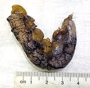
Appendicitis with congestion

Serosa with a patchy purulent exudate.

Appendicitis, with the lumen containing a blood-tinged purulent exudate.

Longitudinal section showing a red inflamed mucosa with an irregular luminal surface (in a case of acute suppurative appendicitis).


Further information: Appendix
Microscopic evaluation
- Evaluate depth of the inflammation.
- Look for any perforation of the wall.
- Look for cancerous cells (which may have caused the appendicitis). Further information: Appendix
- (Attempt to specify the type of appendicitis as either of the following:)
Types
| Pattern | Gross pathology | Light microscopy | Image | Clinical significance |
|---|---|---|---|---|
| Acute intraluminal inflammation | None visible |
|

|
Probably none |
| Acute mucosal inflammation | None visible |
|
May be secondary to enteritis. | |
| Suppurative acute appendicitis | May be inapparent.
|
|

|
Can be presumed to be primary cause of symptoms |
| Gangrenous/necrotizing appendicitis |
|
|

|
Will perforate if untreated |
| Periappendicitis | May be inapparent.
|
|

|
If isolated, probably secondary to other disease |
| Eosinophilic appendicitis | None visible |
|
Possibly parasitic, or eosinophilic enteritis. | |
| Chronic appendicitis[2] |
|
|
Should preferably correlate with long-term or recurrent symptoms. |
Further workup
(In acute suppurative appendicitis, still look for any periappendicitis. Also look by the lumen for parasites.)
Microscopy report
Should include, if detected:
- Acute or chronic appendicitis
- Depth of inflammation
- Any abscess and\or perforation
- Necrosis and\or ulceration, at least if transmural
(Classification into one or several types as per table above.)
- Example
| (Appendix, resection (or appendectomy):) Acute appendicitis and periappendicitis with transmural necrosis and perforation. |
Notes
- ↑ For a full list of contributors, see article history. Creators of images are attributed at the image description pages, seen by clicking on the images. See Patholines:Authorship for details.
Main page
References
- ↑ Unless otherwise specified in rows, reference is:
- Carr, Norman J. (2000). "The pathology of acute appendicitis ". Annals of Diagnostic Pathology 4 (1): 46–58. doi:. ISSN 10929134. - ↑ Sierakowski, Kyra; Pattichis, Andrew; Russell, Patrick; Wattchow, David (2016). "Unusual presentation of a familiar pathology: chronic appendicitis ". BMJ Case Reports: bcr2015212485. doi:. ISSN 1757-790X.
Image sources