Brain/meningeal tumor
Author:
Mikael Häggström [note 1]
Contents
Intraoperative consultation of brain tumor fragments
Preparation
If you are expecting a brain/meningeal tumor, look at any radiology to find out what is the suspected diagnosis or differential diagnoses. A connection to the dura raises the suspicion of a meningioma. Multiple tumors raises the suspicion of metastasis or lymphoma.
Grossing
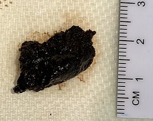
Measure the size of the specimen in 3 dimensions.

- A woven architectural pattern
- Psammoma bodies (spheroid calcifications)
- Syncytial cells (having indistinct cell membranes) with eosinophilic (pink) cytoplasms
- Round uniform nuclei
- Whorls (concentric cell arrangements)[1]
Squash prep
Remove a drop-size sample, place it on a glass-slide, then gently smear it out with another glass slide, followed by applying a fixative solution and staining with H&E.
Evaluation
The most common primary brain tumors are:[2]
- Gliomas[3] (50.4%)
- Meningiomas[3] (20.8%)
- Pituitary adenomas[3] (15%)
- Nerve sheath tumors (10%)
Also look into the patient's history for past cancers that may have metastasized to the brain.
The location, younger vs older age, as well as the overall pattern provide the main differential diagnoses:
Location and younger vs older age
| Location | Child or young adult | Older adult |
|---|---|---|
| Cerebral, supratentorial | Ganglioglioma, dysembryoplastic neuroepithelial tumor (DNET), pleomorphic xanthoastrocytoma (PXA),
ependymoma, atypical teratoid/rhabdoid tumor (AT/RT), CNS embryonal neoplasms |
Glioblastoma, infiltrating astrocytoma (grades II-III), oligodendroglioma, metastasis, lymphoma, infection |
| Cerebellar, infratentorial, fourth ventricle | Pilocytic astrocytoma, medulloblastoma, ependymoma, choroid plexus papilloma, atypical teratoid/rhabdoid tumor (AT/RT) | Metastasis, hemangioblastoma, choroid plexus papilloma, subependymoma |
| Brainstem | Pilocytic astrocytoma, diffuse midline glioma | Astrocytoma, glioblastoma, diffuse midline glioma, metastasis |
| Spinal cord (intramedullary) | Ependymoma, pilocytic astrocytoma, diffuse midline glioma, myxopapillary ependymoma, drop metastasis | Ependymoma, astrocytoma, diffuse midline glioma, myxopapillary ependymoma (filum terminale), paraganglioma (filum terminale) |
| Spinal cord (extramedullary) | Meningioma, schwannoma, metastasis, melanocytoma, melanoma | Schwannoma, meningioma, melanocytoma, melanoma, malignant peripheral nerve sheath tumor (MPNST) |
| Spinal cord (extradural) | Bone tumor, meningioma, abscess, vascular malformation, | Herniated disk, lymphoma, abscess, metastases, |
| Extra-axial, dural, leptomeningeal | Leukemia/lymphoma, Ewing sarcoma, rhabdomyosarcoma, disseminated medulloblastoma, diffuse leptomeningeal glioneuronal tumor (DLGNT), | Meningioma, solitary fibrous tumor, metastasis, lymphoma |
| Sellar/infundibular | Pituitary adenoma, craniopharyngioma, Rathke cleft cyst, pituicytoma, Langerhans cell histiocytosis (LCH), germ cell tumors | Pituitary adenoma, craniopharyngioma, Rathke cleft cyst, pituicytoma, meningioma,
metastasis, chordoma |
| Suprasellar, hypothalamic, optic pathway, third ventricle | Germ cell tumors, craniopharyngioma, pituitary adenoma, optic glioma, Langerhans cell histiocytosis (LCH) | Colloid cyst, craniopharyngioma, chordoid glioma |
| Pineal | Germ cell tumors, pineocytoma, pineoblastoma, pineal cyst | Pineocytoma, pineal cyst, pineal parenchymal tumors of intermediate differentiation (PPTID) |
| Thalamus | Pituitary adenoma, diffuse midline glioma | Diffuse midline glioma, glioblastoma, lymphoma |
| Lateral ventricle | Central neurocytoma, subependymal giant cell astrocytoma (SEGA), choroid plexus papilloma/carcinoma,
meningioma |
Central neurocytoma, subependymal giant cell astrocytoma (SEGA), choroid plexus papilloma/carcinoma, subependymoma, meningioma |
| Nerve root, paraspinal | Neurofibroma, schwannoma, malignant peripheral nerve sheath tumor (MPNST) | Neurofibroma, schwannoma, MPNST, lymphoma, meningioma |
| Cerebellopontine angle | Schwannoma, Choroid plexus papilloma, atypical teratoid/rhabdoid tumor (AT/RT) | Schwannoma, meningioma, epidermoid cyst, choroid plexus papilloma, endolymphatic sac tumor |
Overall patterns
Hypercellular infiltrate but intact architecture:
A sharply demarcated lesion:
Mostly solid, but with ill-defined margin towards adjacent brain tissue:
Extensive necrosis and destruction of normal tissue:
|
Centered around blood vessels:
Outside brain or spine
Only very subtle abnormalities:
Subarachnoid space expansion:
|
Notes
- ↑ For a full list of contributors, see article history. Creators of images are attributed at the image description pages, seen by clicking on the images. See Patholines:Authorship for details.
Main page
References
- ↑ Image by Mikael Häggström, MD. Reference for typical findings: Chunyu Cai, M.D., Ph.D.. Meningioma. Pathology Outlines. Last author update: 10 November 2021}}
- ↑ Park, Bong Jin; Kim, Han Kyu; Sade, Burak; Lee, Joung H. (2009). "Epidemiology". Meningiomas: Diagnosis, Treatment, and Outcome . Springer. p. 11. ISBN 978-1-84882-910-7.
- ↑ 3.0 3.1 3.2 . Brain Tumors - Classifications, Symptoms, Diagnosis and Treatments (in en). www.aans.org.
- ↑ 4.0 4.1 4.2 4.3 4.4 4.5 4.6 4.7 4.8 From notes by Dr. Kurt Schaberg, in turn citing: Perry, Arie; Brat, Daniel J. (2017-12-07). Practical Surgical Neuropathology . Philadelphia, PA: Churchill Livingstone. ISBN 978-0-323-44941-0.
Image sources
