Difference between revisions of "Cirrhosis"
m (→Microscopic evaluation: Format) |
(+Cause) |
||
| Line 4: | Line 4: | ||
}} | }} | ||
Cirrhosis of the [[liver]]: | Cirrhosis of the [[liver]]: | ||
| − | |||
==Gross processing== | ==Gross processing== | ||
In [[autopsy]], make consecutive liver slices, such as in the sagittal or coronal plane. | In [[autopsy]], make consecutive liver slices, such as in the sagittal or coronal plane. | ||
| Line 22: | Line 21: | ||
*'''Diagnosing''' cirrhosis. The diagnosis of cirrhosis by biopsy requires the presence of fibrosis and nodules. | *'''Diagnosing''' cirrhosis. The diagnosis of cirrhosis by biopsy requires the presence of fibrosis and nodules. | ||
*Assessing the degree of '''inflammation''' (grade) and '''fibrosis''' (stage) of the disease. | *Assessing the degree of '''inflammation''' (grade) and '''fibrosis''' (stage) of the disease. | ||
| − | + | *Finding the most probable differential diagnoses that '''cause''' the cirrhosis (or correlating with a previously established diagnosis). | |
<gallery mode=packed heights=180> | <gallery mode=packed heights=180> | ||
File:Histopathology of mild zone 3 steatosis without fibrosis (van Gieson).jpg|'''No fibrosis''', but mild zone 3 steatosis, in which collagen fibres (pink–red, arrow) are confined to portal tracts (P) (van Gieson's stain)<ref name="BoydCain2020">{{cite journal|last1=Boyd|first1=Alexander|last2=Cain|first2=Owen|last3=Chauhan|first3=Abhishek|last4=Webb|first4=Gwilym James|title=Medical liver biopsy: background, indications, procedure and histopathology|journal=Frontline Gastroenterology|volume=11|issue=1|year=2020|pages=40–47|issn=2041-4137|doi=10.1136/flgastro-2018-101139}}<br>-"This is an open access article distributed in accordance with the Creative Commons Attribution 4.0 Unported (CC BY 4.0) license"</ref> | File:Histopathology of mild zone 3 steatosis without fibrosis (van Gieson).jpg|'''No fibrosis''', but mild zone 3 steatosis, in which collagen fibres (pink–red, arrow) are confined to portal tracts (P) (van Gieson's stain)<ref name="BoydCain2020">{{cite journal|last1=Boyd|first1=Alexander|last2=Cain|first2=Owen|last3=Chauhan|first3=Abhishek|last4=Webb|first4=Gwilym James|title=Medical liver biopsy: background, indications, procedure and histopathology|journal=Frontline Gastroenterology|volume=11|issue=1|year=2020|pages=40–47|issn=2041-4137|doi=10.1136/flgastro-2018-101139}}<br>-"This is an open access article distributed in accordance with the Creative Commons Attribution 4.0 Unported (CC BY 4.0) license"</ref> | ||
Revision as of 12:15, 31 March 2023
Author:
Mikael Häggström [note 1]
Cirrhosis of the liver:
Contents
Gross processing
In autopsy, make consecutive liver slices, such as in the sagittal or coronal plane.
Gross examination
Cirrhosis is characterized by pale areas. Categorize into either of the following: (1) micronodular, (2) macronodular, or (3) mixed:[1]

- Micronodular or diffuse cirrhosis (uniform nodules less than 3 mm in diameter).[notes 1]
- Macronodular cirrhosis (irregular nodules with a variation greater than 3 mm in diameter).
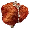
Pale macronodules of cirrhosis
.jpg)
- Mixed cirrhosis (when features of both micronodular and macronodular cirrhosis are present): Usually micronodular cirrhosis progresses into macronodular cirrhosis over time.
Microscopic evaluation
Components:[1]
- Diagnosing cirrhosis. The diagnosis of cirrhosis by biopsy requires the presence of fibrosis and nodules.
- Assessing the degree of inflammation (grade) and fibrosis (stage) of the disease.
- Finding the most probable differential diagnoses that cause the cirrhosis (or correlating with a previously established diagnosis).

No fibrosis, but mild zone 3 steatosis, in which collagen fibres (pink–red, arrow) are confined to portal tracts (P) (van Gieson's stain)[2]

Histopathology of steatohepatitis with mild fibrosis in the form of fibrous expansion (van Gieson's stain)[2]

Histopathology of steatohepatitis with moderate fibrosis, with thin fibrous bridges (van Gieson's stain)[2]

Moderate fibrosis (H&E stain).

Histopathology of steatohepatitis with established cirrhosis, with thick bands of fibrosis (van Gieson's stain)[2]

Trichrome stain, showing cirrhosis as a nodular texture surrounded by fibrosis (wherein collagen is stained blue).
.jpg)
.jpg)
.jpg)

.jpg)
_(5690946257).jpg)
Notes
- ↑ Micronodular or diffuse cirrhosis can be due to alcohol, hemochromatosis, hepatic venous outflow obstruction, chronic biliary obstruction, jejunoileal bypass, and Indian childhood cirrhosis.
- ↑ For a full list of contributors, see article history. Creators of images are attributed at the image description pages, seen by clicking on the images. See Patholines:Authorship for details.
Main page
References
- ↑ 1.0 1.1 Bashar Sharma; Savio John.. Hepatic Cirrhosis. StatPearls at the National Center for Biotechnology Information. Last Update: June 3, 2019.
- ↑ 2.0 2.1 2.2 2.3 Boyd, Alexander; Cain, Owen; Chauhan, Abhishek; Webb, Gwilym James (2020). "Medical liver biopsy: background, indications, procedure and histopathology
". Frontline Gastroenterology 11 (1): 40–47. doi:. ISSN 2041-4137.
-"This is an open access article distributed in accordance with the Creative Commons Attribution 4.0 Unported (CC BY 4.0) license"
Image sources