Difference between revisions of "Colorectal carcinoma"
Jump to navigation
Jump to search
m (despaced) |
(Templated) |
||
| Line 10: | Line 10: | ||
==Microscopic evaluation== | ==Microscopic evaluation== | ||
{{Colorectal adenocarcinoma - microscopy criteria}} | {{Colorectal adenocarcinoma - microscopy criteria}} | ||
| + | |||
| + | ===Differential diagnosis=== | ||
| + | {{Colorectal adenoma versus adenocarcinoma}} | ||
| + | |||
===Staging=== | ===Staging=== | ||
Determine depth of growth and/or infiltration. Preferably stage by the AJCC or TNM system: | Determine depth of growth and/or infiltration. Preferably stage by the AJCC or TNM system: | ||
{{Colorectal cancer staging }} | {{Colorectal cancer staging }} | ||
{{Bottom}} | {{Bottom}} | ||
Revision as of 04:29, 30 September 2019
Author:
Mikael Häggström [note 1]
Contents
Gross evaluation
Depending on presentation:
Microscopic evaluation
Microscopy criteria for colorectal adenocarcinoma
- A lesion at least "high grade intramucosal neoplasia" (high grade dysplasia) has:
- Severe cytologic atypia[1]
- Cribriform architecture, consisting of juxtaposed gland lumens without stroma in between, with loss of cell polarity. Rarely, they have foci of squamous differentiation (morules).[1]
- This should be distinguished from cases where piles of well-differentiated mucin-producing cells appear cribriform. In such piles, nuclei show regular polarity with apical mucin, and their nuclei are not markedly enlarged.[1]
- Invasive adenocarcinoma commonly displays:
- Varying degrees of gland formation with tall columnar cells.[1]
- Frequenty desmoplasia.[1]
- Dirty necrosis, consisting of extensive central necrosis with granular eosinophilic karyorrhectic cell detritus.[1][2] It is located within the glandular lumina,[2] or often with a garland of cribriform glands in their vicinity.[1]
It may also show lymphovascular invasion.

Desmoplastic reaction (*)
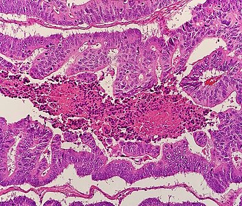
Dirty necrosis

Lymphovascular invasion (lymphatic invasion pictured)


Differential diagnosis
Colorectal carcinoma (mainly adenocarcinoma) is distinguished from an adenoma (mainly tubular and ⁄or villous adenomas) mainly by invasion through the muscularis mucosae.[3]
Staging
Determine depth of growth and/or infiltration. Preferably stage by the AJCC or TNM system:
| AJCC stage[4] | TNM stage[4] | TNM stage criteria[4] |
|---|---|---|
| Stage 0 | Tis N0 M0 | Tis: Tumor confined to mucosa; cancer-in-situ |
| Stage I | T1 N0 M0 | T1: Tumor invades submucosa |
| T2 N0 M0 | T2: Tumor invades muscularis propria | |
| Stage II-A | T3 N0 M0 | T3: Tumor invades subserosa or beyond (without other organs involved) |
| Stage II-B | T4a N0 M0 | T4a: Tumor perforates the visceral peritoneum |
| Stage II-C | T4b N0 M0 | T4b: Tumor invades adjacent organs |
| Stage III-A |
|
|
| Stage III-B |
|
|
| Stage III-C |
|
|
| Stage IVa | any T, any N, M1a | M1a: Metastasis to 1 other part of the body beyond the colon, rectum or regional lymph nodes. Any T, any N. |
| Stage IVb | any T, any N, M1b | M1b: Metastasis to more than 1 other part of the body beyond the colon, rectum or regional lymph nodes. Any T, any N. |
| Stage IVc | any T, any N, M1c | M1c: Metastasis to the peritoneal surface. Any T, any N. |
Notes
- ↑ For a full list of contributors, see article history. Creators of images are attributed at the image description pages, seen by clicking on the images. See Patholines:Authorship for details.
Main page
References
- ↑ 1.0 1.1 1.2 1.3 1.4 1.5 1.6 Robert V Rouse. Adenocarcinoma of the Colon and Rectum. Stanford University School of Medicine. Original posting/updates: 1/31/10, 7/15/11, 11/12/11
- ↑ 2.0 2.1 Li, Lianhuang; Jiang, Weizhong; Yang, Yinghong; Chen, Zhifen; Feng, Changyin; Li, Hongsheng; Guan, Guoxian; Chen, Jianxin (2014). "Identification of dirty necrosis in colorectal carcinoma based on multiphoton microscopy ". Journal of Biomedical Optics 19 (6): 066008. doi:. ISSN 1083-3668.
- ↑ Robert V Rouse. Colorectal Adenoma Containing Invasive Adenocarcinoma. Stanford University School of Medicine.
- ↑ 4.0 4.1 4.2 . Colorectal Cancer: Stages. Cancer.net (American Society of Clinical Oncology). Retrieved on 2019-09-26. Approved by the Cancer.Net Editorial Board, 11/2018. In turn citing:
Amin, Mahul B.; Greene, Frederick L.; Edge, Stephen B.; Compton, Carolyn C.; Gershenwald, Jeffrey E.; Brookland, Robert K.; Meyer, Laura; Gress, Donna M.; et al. (2017). "The Eighth Edition AJCC Cancer Staging Manual: Continuing to build a bridge from a population-based to a more “personalized” approach to cancer staging ". CA: A Cancer Journal for Clinicians 67 (2): 93–99. doi:. ISSN 00079235.
Image sources