Difference between revisions of "Evaluation"
Jump to navigation
Jump to search
m (→Artifacts: delinked) |
(+History) |
||
| Line 5: | Line 5: | ||
[[File:Position of objective (edited).jpg|thumb|210px|Example starting position of objective.]] | [[File:Position of objective (edited).jpg|thumb|210px|Example starting position of objective.]] | ||
[[File:Microscopy slide scanning.jpg|thumb|Example slide scanning directions.]] | [[File:Microscopy slide scanning.jpg|thumb|Example slide scanning directions.]] | ||
| − | * | + | *Preferably, look up past '''medical history''' of the patient, mainly past cancers that could possibly appear in the current specimen. |
| + | *Look at each microscopy slide by '''eye''', to plan the microscopy screening so as to not miss peripheral fragments. | ||
*Have a systematic '''direction''' of screening through microscopy slides, such as from top left to bottom right as seen in the microscope. When two-way mirrored, the starting position of the microscope slide is then with the objective pointing at bottom right. | *Have a systematic '''direction''' of screening through microscopy slides, such as from top left to bottom right as seen in the microscope. When two-way mirrored, the starting position of the microscope slide is then with the objective pointing at bottom right. | ||
Revision as of 14:41, 3 August 2021
Author:
Mikael Häggström [note 1]
- Preferably, look up past medical history of the patient, mainly past cancers that could possibly appear in the current specimen.
- Look at each microscopy slide by eye, to plan the microscopy screening so as to not miss peripheral fragments.
- Have a systematic direction of screening through microscopy slides, such as from top left to bottom right as seen in the microscope. When two-way mirrored, the starting position of the microscope slide is then with the objective pointing at bottom right.
Artifacts
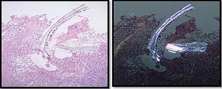
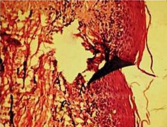
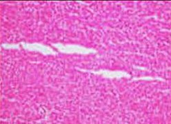
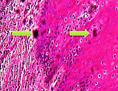
In microscopy, an artifact is an apparent structural detail that is caused by the processing of the specimen and is thus not a legitimate feature of the specimen. Major artifacts to account for include:
Notes
- ↑ For a full list of contributors, see article history. Creators of images are attributed at the image description pages, seen by clicking on the images. See Patholines:Authorship for details.
Main page
References
Image sources


