Fundic gland polyp
Author:
Mikael Häggström [note 1]
Contents
Presentation
- Mainly gastric polyp.
Microscopic evaluation

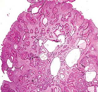
A fundic gland polyp displays cystically dilated glands lined by chief cells and parietal cells, and possibly also mucinous foveolar cells.[1]

A fundic gland polyp, low magnification.[image 1]


Differential diagnosis
Gastric hyperplastic polyp: Elongated, tortuous, and cystic foveolae separated by edematous and inflamed stroma.[2]

Gastric hyperplastic polyp, high magnification, showing the edematous and inflamed stroma.


Workup

Look for dysplasia (center), compared to normal mucosa (at right). [image 1]

Dysplasia in fundic gland polyp is mainly seen as nuclear enlargement, hyperchromasia, pseudostratification, and loss of cytoplasmic mucin.[3] However, you don't need to spend much effort in the decision, since these patients have an excellent prognosis whether there is dysplasia or not.
Reporting
Generally, keep it short:
| (Stomach, excision:) Fundic gland polyp |
Notes
- ↑ For a full list of contributors, see article history. Creators of images are attributed at the image description pages, seen by clicking on the images. See Patholines:Authorship for details.
Main page
References
- ↑ Naziheh Assarzadegan, M.D., Raul S. Gonzalez, M.D.. Stomach Polyps - Fundic gland polyp. PathologyOutlines. Topic Completed: 1 November 2017. Minor changes: 11 December 2019
- ↑ Groisman, Gabriel M.; Depsames, Roman; Ovadia, Baruch; Meir, Alona (2014). "Metastatic Carcinoma Occurring in a Gastric Hyperplastic Polyp Mimicking Primary Gastric Cancer: The First Reported Case
". Case Reports in Pathology 2014: 1–5. doi:. ISSN 2090-6781.
- Attribution 3.0 Unported (CC BY 3.0) license - ↑ Levy MD, Bhattacharya B (2015). "Sporadic Fundic Gland Polyps With Low-Grade Dysplasia: A Large Case Series Evaluating Pathologic and Immunohistochemical Findings and Clinical Behavior. ". Am J Clin Pathol 144 (4): 592-600. doi:. PMID 26386080. Archived from the original. .
Image sources
- ↑ 1.0 1.1 Image(s) by: Mikael Häggström, M.D. Public Domain
- Author info
- Reusing images