Difference between revisions of "Gastric polyp"
Jump to navigation
Jump to search

(Templated) |
(Templated) |
||
| Line 3: | Line 3: | ||
|author2= | |author2= | ||
}} | }} | ||
| + | {{Endoscopic biopsies}} | ||
==Microscopic evaluation== | ==Microscopic evaluation== | ||
[[File:Pie chart of relative incidences of gastric polyps.png|thumb|Relative incidences of gastric polyps. The remaining 4.8% are mainly constituted by lipomas, GIST, xanthomas and inflammatory pseudopolyps.<ref>{{cite journal|last1=García-Alonso|first1=Francisco Javier|last2=Martín-Mateos|first2=Rosa María|last3=González-Martín|first3=Juan Ángel|last4=Foruny|first4=José Ramón|last5=Vázquez-Sequeiros|first5=Enrique|last6=Boixeda de Miquel|first6=Daniel|title=Gastric polyps: analysis of endoscopic and histological features in our center|journal=Revista Española de Enfermedades Digestivas|volume=103|issue=8|year=2011|pages=416–420|issn=1130-0108|doi=10.4321/S1130-01082011000800005}}</ref>]] | [[File:Pie chart of relative incidences of gastric polyps.png|thumb|Relative incidences of gastric polyps. The remaining 4.8% are mainly constituted by lipomas, GIST, xanthomas and inflammatory pseudopolyps.<ref>{{cite journal|last1=García-Alonso|first1=Francisco Javier|last2=Martín-Mateos|first2=Rosa María|last3=González-Martín|first3=Juan Ángel|last4=Foruny|first4=José Ramón|last5=Vázquez-Sequeiros|first5=Enrique|last6=Boixeda de Miquel|first6=Daniel|title=Gastric polyps: analysis of endoscopic and histological features in our center|journal=Revista Española de Enfermedades Digestivas|volume=103|issue=8|year=2011|pages=416–420|issn=1130-0108|doi=10.4321/S1130-01082011000800005}}</ref>]] | ||
Revision as of 10:25, 16 November 2020
Author:
Mikael Häggström [note 1]
| Mostly: |
Microscopic evaluation

Relative incidences of gastric polyps. The remaining 4.8% are mainly constituted by lipomas, GIST, xanthomas and inflammatory pseudopolyps.[1]
Gastric hyperplastic polyp: Elongated, tortuous, and cystic foveolae separated by edematous and inflamed stroma.[2]

Gastric hyperplastic polyp, high magnification

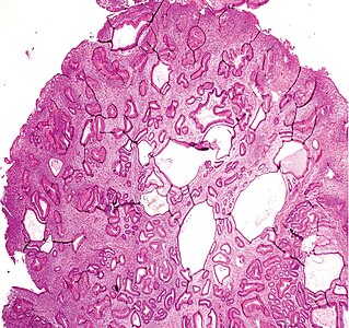
A fundic gland polyp displays cystically dilated glands lined by chief cells and parietal cells, and possibly also mucinous foveolar cells.[3]

A fundic gland polyp, low magnification.[image 1]


Notes
- ↑ For a full list of contributors, see article history. Creators of images are attributed at the image description pages, seen by clicking on the images. See Patholines:Authorship for details.
Main page
References
- ↑ García-Alonso, Francisco Javier; Martín-Mateos, Rosa María; González-Martín, Juan Ángel; Foruny, José Ramón; Vázquez-Sequeiros, Enrique; Boixeda de Miquel, Daniel (2011). "Gastric polyps: analysis of endoscopic and histological features in our center ". Revista Española de Enfermedades Digestivas 103 (8): 416–420. doi:. ISSN 1130-0108.
- ↑ Groisman, Gabriel M.; Depsames, Roman; Ovadia, Baruch; Meir, Alona (2014). "Metastatic Carcinoma Occurring in a Gastric Hyperplastic Polyp Mimicking Primary Gastric Cancer: The First Reported Case
". Case Reports in Pathology 2014: 1–5. doi:. ISSN 2090-6781.
- Attribution 3.0 Unported (CC BY 3.0) license - ↑ Naziheh Assarzadegan, M.D., Raul S. Gonzalez, M.D.. Stomach Polyps - Fundic gland polyp. PathologyOutlines. Topic Completed: 1 November 2017. Minor changes: 11 December 2019
Image sources
- ↑ Image(s) by: Mikael Häggström, M.D. Public Domain
- Author info
- Reusing images