Granulomatous skin inflammation
Jump to navigation
Jump to search
Author:
Mikael Häggström [note 1]
Contents
Microscopic examination
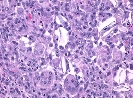
Granulomatous inflammation is defined by the presence of mononuclear leukocytes, specifically histiocytes, appearing as epithelioid cells with round to oval nuclei, often with irregular contours and abundant granular eosinophilic cytoplasm with indistinct cell borders. They may also coalesce to form multinucleated giant cells.[1]
Differential diagnosis
- Other forms of dermatitis if other types of inflammatory cells are prevalent.
- Suspected malignant skin lesions if demonstrating cellular atypia
Workup
- Look for foreign bodies, conferring a diagnosis of foreign body granuloma.
- Look for any specific appearance of the giant cells.
- Look for relation to adnexae or other particular structures.

Foreign body: Suture granuloma, with multinucleated giant cells surrounding (grey) suture material.

Relation: Folliculitis with multinucleated giant cells surrounding a hair follicle.
Granulomatious inflammation following an epidermal inclusion cyst rupture, with giant cells surrounding keratin flakes.

.jpg)


Notes
- ↑ For a full list of contributors, see article history. Creators of images are attributed at the image description pages, seen by clicking on the images. See Patholines:Authorship for details.
Main page
References
- ↑ Shah, Kabeer K.; Pritt, Bobbi S.; Alexander, Mariam P. (2017). "Histopathologic review of granulomatous inflammation ". Journal of Clinical Tuberculosis and Other Mycobacterial Diseases 7: 1–12. doi:. ISSN 24055794.
- ↑ Grant-Kels, Jane (2007). Color Atlas of Dermatopathology . City: Informa Healthcare. pp. 107, 119. ISBN 978-0-8493-3794-9.
- ↑ Carmen Gómez-Mateo, Maria; Monteagudo, Carlos (2013). "Nonepithelial skin tumors with multinucleated giant cells ". Seminars in Diagnostic Pathology 30 (1): 58–72. doi:. PMID 23327730. Archived from the original. .
- ↑ Sequeira, Fiona; Gandhi, Suneil (2012). "Named cells in dermatology ". Indian Journal of Dermatology, Venereology and Leprology 78 (2): 207–16. doi:. PMID 22421663. Archived from the original. .
Image sources