Difference between revisions of "Hepatocellular carcinoma"
Jump to navigation
Jump to search

(→Microscopic evaluation: +Adenoma) |
(→Differentiation: +Gallery) |
||
| Line 28: | Line 28: | ||
===Differentiation=== | ===Differentiation=== | ||
Differentiations of hepatocellular carcinoma are:<ref name=Deepali/> | Differentiations of hepatocellular carcinoma are:<ref name=Deepali/> | ||
| − | |||
*'''Well differentiated''': | *'''Well differentiated''': | ||
:*Thin plates (1 - 3 hepatocytes thick) | :*Thin plates (1 - 3 hepatocytes thick) | ||
| Line 35: | Line 34: | ||
:*Minimal nuclear atypia | :*Minimal nuclear atypia | ||
:*Nuclear density of 2x compared to normal liver | :*Nuclear density of 2x compared to normal liver | ||
| − | |||
*'''Moderately differentiated''': | *'''Moderately differentiated''': | ||
:*Trabecular pattern at least 4 cells thick | :*Trabecular pattern at least 4 cells thick | ||
| Line 41: | Line 39: | ||
:*Distinct nucleoli | :*Distinct nucleoli | ||
:*Pseudoglands | :*Pseudoglands | ||
| − | |||
*'''Poorly differentiated''': | *'''Poorly differentiated''': | ||
:*Large tumor cells with hyperchromatic nuclei in compact growth pattern | :*Large tumor cells with hyperchromatic nuclei in compact growth pattern | ||
| Line 47: | Line 44: | ||
:*Prominent pleomorphism | :*Prominent pleomorphism | ||
:*May have spindle cell or small cell areas | :*May have spindle cell or small cell areas | ||
| + | <gallery mode=packed heights=190> | ||
| + | File:Histopathology of well-differentiated hepatocellular carcinoma.jpg|Well-differentiated HCC. | ||
| + | File:Histopathology of moderately differentiated hepatocellular carcinoma.jpg|Moderately differentiated HCC. | ||
| + | File:Histopathology of poorly differentiated hepatocellular carcinoma.jpg|Poorly differentiated HCC. | ||
| + | </gallery> | ||
{{Bottom}} | {{Bottom}} | ||
Revision as of 17:24, 22 February 2021
Author:
Mikael Häggström [note 1]
Contents
Presentations
Hepatocellular carcinoma is the most common diagnosis for liver tumors.:[1]
Microscopic evaluation
Hepatocellular carcinoma typically displays:[2]
- Trabecular pattern surrounded by layer of flattened endothelial cells.
- Presence of sinusoidal vessels surrounding tumor cells
- Scant stroma, and minimal desmoplasia
- Polygonal cells with distinct cell membranes
- Abundant granular eosinophilic cytoplasm
- Nuclei with increased N/C ratio, round nuclei with coarse chromatin and thickened nuclear membrane, and may have prominent nucleoli.
Needle biopsy.

Needle biopsy.

Needle biopsy.

FNA.

FNA clot section.

FNA clot section.
.jpg)
.jpg)
.jpg)
.jpg)
.jpg)
.jpg)
Hepatocellular adenoma versus carcinoma

Histopathology of inflammatory hepatocellular adenoma, with inflammatory cells and minor atypia.[3]
A hepatocellular adenoma, in contrast to a carcinoma, has a well-defined border between the lesion and background liver, and is composed of hepatocytes with less significant cytologic atypia.[4]
Both hepatocellular adenoma and well-differentiated hepatocellular carcinoma may display:
- Steatosis: A steatotic hepatocellular carcinoma is a common variant, found most commonly in small, well-differentiated tumors.[5]
- Portal triads, which commonly persist in well-differentiated hepatocellular carcinomas.[6]
Differentiation
Differentiations of hepatocellular carcinoma are:[2]
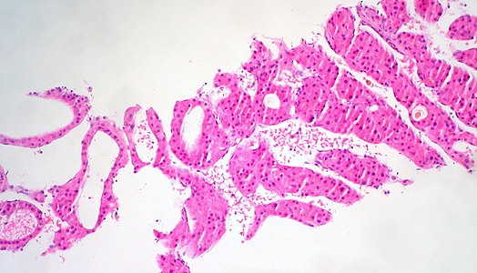
- Well differentiated:
- Thin plates (1 - 3 hepatocytes thick)
- Hepatocytes are smaller than normal
- Abnormal reticulin network
- Minimal nuclear atypia
- Nuclear density of 2x compared to normal liver
- Moderately differentiated:
- Trabecular pattern at least 4 cells thick
- Larger cells with more eosinophilic cytoplasm
- Distinct nucleoli
- Pseudoglands
- Poorly differentiated:
- Large tumor cells with hyperchromatic nuclei in compact growth pattern
- Rare trabeculae or bile
- Prominent pleomorphism
- May have spindle cell or small cell areas

Well-differentiated HCC.

Moderately differentiated HCC.

Poorly differentiated HCC.


Notes
- ↑ For a full list of contributors, see article history. Creators of images are attributed at the image description pages, seen by clicking on the images. See Patholines:Authorship for details.
Main page
References
- ↑ Table 37.2 in: Sternberg, Stephen (2012). Sternberg's diagnostic surgical pathology . Place of publication not identified: LWW. ISBN 978-1-4511-5289-0. OCLC 953861627.
- ↑ 2.0 2.1 Deepali Jain. Liver & intrahepatic bile ducts - Hepatocellular carcinoma - Hepatocellular carcinoma overview. PathologyOutlines. Topic Completed: 1 February 2012. Minor changes: 30 September 2020
- ↑ Bioulac-Sage, Paulette; Sempoux, Christine; Possenti, Laurent; Frulio, Nora; Laumonier, Hervé; Laurent, Christophe; Chiche, Laurence; Frédéric Blanc, Jean; et al. (2013). "Pathological Diagnosis of Hepatocellular Cellular Adenoma according to the Clinical Context ". International Journal of Hepatology 2013: 1–13. doi:. ISSN 2090-3448.
- ↑ Author: Avani Pendse, M.D., Ph.D.. Liver & intrahepatic bile ducts - Benign / nonneoplastic - Hepatocellular adenoma. Pathology Outlines. Topic Completed: 1 October 2018. Minor changes: 30 September 2020
- ↑ Chan, Anthony W H; Yu, Shuangni; Yu, Yau-Hei; Tong, Joanna H M; Wang, Lei; Tin, Edith K Y; Chong, Charing C N; Chan, Stephen L; et al. (2016). "Steatotic hepatocellular carcinoma: a variant associated with metabolic factors and late tumour relapse ". Histopathology 69 (6): 971–984. doi:. ISSN 03090167.
- ↑ Motohashi, Ikuko; Okudaira, Masahiko; Takai, Tomoko; Kaneko, Satoshi; Ikeda, Noriaki (1992). "Morphological differences between hepatocellular carcinoma and hepatocellular carcinomalike lesions ". Hepatology 16 (1): 118–126. doi:. ISSN 02709139.
Image sources