Difference between revisions of "Hysterectomy"
Jump to navigation
Jump to search
(→Gross processing: +Indications) |
(→Microscopic evaluation: +Findings) |
||
| Line 51: | Line 51: | ||
===Cervix=== | ===Cervix=== | ||
{{CIN grades}} | {{CIN grades}} | ||
| + | |||
| + | ===Uterine body=== | ||
| + | Main findings: | ||
| + | <gallery mode=packed heights=200> | ||
| + | File:Histopathology of adenomyosis.jpg|Adenomyosis (endometrial glands in the myometrium) | ||
| + | File:Histopathology of uterine leiomyoma.jpg|'''[[Smooth muscle tumor]]''' (usually whorled pattern) | ||
| + | </gallery> | ||
{{Bottom}} | {{Bottom}} | ||
Revision as of 10:53, 17 March 2020
Author:
Mikael Häggström [note 1]
Contents
Fixation
Generally 10% neutral buffered formalin.
See also: General notes on fixation
Gross processing
Benign indications
Applicable in bleeding disorders, pain, leiomyoma and endometrial hyperplasia.[1]
Gross examination
For orientation:
- The round ligament lies anterior to the tubes and ovaries.[1]
- The peritoneum extends further down along the cervix posteriorly than anteriorly.[2] Its ends bluntly posteriorly and sharply anteriorly.[2]
Optionally, remove the adnexa.[1]
Steps:[1]
- Perform a general inspection
- Measure length, width, thickness
- The uterus is usually opened at the front in the midline, optionally with an incision towards each corner.[notes 1] Open the cavity completely, along the existing incision. The cavity is sometimes be squeezed or rolled around a leiomyoma, and you'll you have to improvise and perhaps go around the leiomyoma to open the cavity properly. Cut through the front wall into both corners.
- Inspect the mucosa. If any polyps: Further information: Endometrial polyp
- Measure the thickness of the mucosa and myometrium

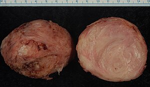
Smooth muscle tumor (in this case leiomyoma).
- Inspect the myometrium. If any tumor: Further information: Smooth muscle tumor
Gross report
Components:[1]
- Shape of uterus and adnex
- Measurements
- Mucosa, such as smooth or irregular.
- Any polyps. Further information: Endometrial polyp
- Mucosal and endometrial thickness
- Any smooth muscle tumor. Further information: Smooth muscle tumor
Slices for microscopy
Submit:[1]
- Four cross-sectiosn from any accompanying ectocervix. In subtotal extirpation, a cross-section is taken from the lower resection border.
- A transverse slice through the endocervix, possibly divided into two.
- One slice from the front and one from the back wall of the corpus, and one piece from each corner, including myometrium.
- Any mucosal parts with macroscopically abnormal appearance, including polyps.
- In case of endometrial hyperplasia, most of the mucosa of the corpus and fundus.
- Samples form all smooth muscle tumors >5 cm in diameter. Further information: Smooth muscle tumor
Microscopic evaluation
Look for signs of malignancy:
- Myometrium tumors: Further information: Smooth muscle tumor
- Endometrial polyps: Further information: Endometrial polyp
- Endometrial hyperplasia: Further information: Endometrial hyperplasia
Cervix
Look for cervical dysplasia. It is mainly seen as nuclei with hyperchromasia, coarse chromatin and irregular contours.[3]

Spectrum from normal to high grade SIL.[4]

Further information: Cervical dysplasia
Uterine body
Main findings:

Adenomyosis (endometrial glands in the myometrium)

Smooth muscle tumor (usually whorled pattern)


Notes
- ↑ The uterus can also be opened laterally, through the parameters.
- ↑ For a full list of contributors, see article history. Creators of images are attributed at the image description pages, seen by clicking on the images. See Patholines:Authorship for details.
Main page
References
- ↑ 1.0 1.1 1.2 1.3 1.4 1.5 Monica Dahlgren, Janne Malina, Anna Måsbäck, Otto Ljungberg. Stora utskärningen. KVAST (Swedish Society of Pathology). Retrieved on 2019-09-26.
- ↑ 2.0 2.1 . General Specimen Orientation Tips. The University of Michigan (2020-01-29).
- ↑ Khaled J. Alkhateeb, M.B.B.S., Ziyan T. Salih, M.D.. HSIL / CIN II / CIN III. PathologyOutlines. Topic Completed: 29 March 2021. Minor changes: 9 February 2022
- ↑ Source image by Ed Uthman from Houston, TX, USA. Creative Commons Attribution 2.0 Generic (CC BY 2.0) license
Image sources