Difference between revisions of "Lipomatous tumor"
m (→Microscopic evaluation: Expanded) |
|||
| Line 33: | Line 33: | ||
For lipomas: Absence of signs of malignancy. | For lipomas: Absence of signs of malignancy. | ||
{|class="wikitable" | {|class="wikitable" | ||
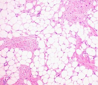
| − | [[File:Histopathology of lipoma.jpg|190px|left]] | + | | [[File:Histopathology of lipoma.jpg|190px|left]] |
Tissue composed of univacuolar fat cells and delicate and inconspicuous fibrous septa. No evidence of malignancy. | Tissue composed of univacuolar fat cells and delicate and inconspicuous fibrous septa. No evidence of malignancy. | ||
|} | |} | ||
{{Reporting}} | {{Reporting}} | ||
{{Bottom}} | {{Bottom}} | ||
Revision as of 11:42, 10 February 2020
Author:
Mikael Häggström [note 1]

Contents
Fixation
Generally 10% neutral buffered formalin.
See also: General notes on fixation
Gross processing
- Perform consecutive slicing of the entire specimen.
- Look for signs of liposarcoma: Mainly by firm volumes.[1] Color varies from yellow to white (and firm) depending on the proportion of adipocytic, fibrous and/or myxoid content.[2] Areas of fat necrosis are common in larger lesions. Rarely, infiltrative growth is seen.[2]
- Submit slices from any suspicious parts, or at least one representative slice from the specimen.

Cross-section of lipoma. Homogenous texture.

Liposarcoma: Tumor section reveals brownish-yellowish areas, hemorrhagic and calcific zones.


Gross report
- Color
- Even absence of hemorrhage or necrosis.
Example:
| Mass weighing 121 grams and measuring 10 x 6,5 x 3,5 cm. Homogenous yellow color. No hemorrhage or necrosis. |
See also: General notes on gross processing
Microscopic evaluation
Lipoma: lobules of mature white adipose tissue divided by delicate and inconspicuous fibrous septa containing thin-walled capillary-sized vessels.

Fibrolipoma: A lipoma with focal areas of large amounts of fibrous tissue. A sclerotic lipoma is one step further: a predominantly fibrous lesion with focal areas of fat.[3]

Myxoid liposarcoma: Hypercellular solid sheets of cells lying back to back, with round cells or primitive cytomorphology.[4]

.jpg)
Microscopy/Histopathology report
For lipomas: Absence of signs of malignancy.
 Tissue composed of univacuolar fat cells and delicate and inconspicuous fibrous septa. No evidence of malignancy. |
See also: General notes on reporting
Notes
- ↑ For a full list of contributors, see article history. Creators of images are attributed at the image description pages, seen by clicking on the images. See Patholines:Authorship for details.
Main page
References
- ↑ Monica Dahlgren, Janne Malina, Anna Måsbäck, Otto Ljungberg (1997-02-13). Lilla utskärningen.
- ↑ 2.0 2.1 Andreas F Mavrogenis, Panayiotis J Papagelopoulos (2013-02-01). Soft Tissues: Well-differentiated liposarcoma. Atlas of Genetics and Cytogenetics in Oncology and Haematology.
- ↑ . Lipoma Variant: Fibrolipoma. Stanford University School of Medicine. Retrieved on 2020-02-10.
- ↑ Michael R. Clay. Soft tissue - Adipose tissue - Myxoid liposarcoma. PathologyOutlines. Topic Completed: 1 January 2018. Revised: 20 March 2019
Image sources