Difference between revisions of "Lipomatous tumor"
(→Microscopic evaluation: Another) |
(→Microscopic evaluation: Essentially) |
||
| Line 29: | Line 29: | ||
<gallery mode=packed heights=220> | <gallery mode=packed heights=220> | ||
File:Histopathology of lipoma.jpg|'''Lipoma''': lobules of mature white adipose tissue divided by delicate and inconspicuous fibrous septa containing thin-walled capillary-sized vessels. Still, look close at the fibrous septa: | File:Histopathology of lipoma.jpg|'''Lipoma''': lobules of mature white adipose tissue divided by delicate and inconspicuous fibrous septa containing thin-walled capillary-sized vessels. Still, look close at the fibrous septa: | ||
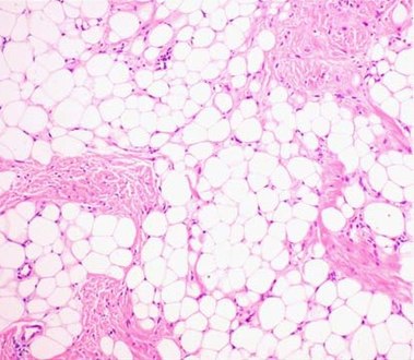
| − | File:Histopathology of an atypical lipomatous tumor or well-differentiated liposarcoma, lipoma-like subtype.jpg|An '''atypical lipomatous tumor''' (also termed '''well-differentiated liposarcoma'''), lipoma-like subtype. At low magnification, the majority of the tumor has the look of benign mature adipocytes, but high magnification of a fibrous band shows spindle cells with enlarged, hyperchromatic nuclei. Another clue for liposarcoma is a higher variability of lipid droplet sizes. | + | File:Histopathology of an atypical lipomatous tumor or well-differentiated liposarcoma, lipoma-like subtype.jpg|An '''atypical lipomatous tumor''' (also termed '''well-differentiated liposarcoma'''), lipoma-like subtype. At low magnification, the majority of the tumor essentially has the look of benign mature adipocytes (except for mildly increased variation in lipid droplet sizes), but high magnification of a fibrous band shows spindle cells with enlarged, hyperchromatic nuclei. Another clue for liposarcoma is a higher variability of lipid droplet sizes. |
File:Histopathology of a fibrolipoma.jpg|'''Fibrolipoma''' is a lipoma with focal areas of large amounts of fibrous tissue.<br>A '''sclerotic lipoma''' is one step further: a predominantly fibrous lesion with focal areas of fat.<ref>{{cite web|url=http://surgpathcriteria.stanford.edu/softfat/lipoma/fibrolipoma.html|title=Lipoma Variant: Fibrolipoma|website=Stanford University School of Medicine|accessdate=2020-02-10}}</ref><br>If unsure of degree of fibrosis: Simply report as lipoma. | File:Histopathology of a fibrolipoma.jpg|'''Fibrolipoma''' is a lipoma with focal areas of large amounts of fibrous tissue.<br>A '''sclerotic lipoma''' is one step further: a predominantly fibrous lesion with focal areas of fat.<ref>{{cite web|url=http://surgpathcriteria.stanford.edu/softfat/lipoma/fibrolipoma.html|title=Lipoma Variant: Fibrolipoma|website=Stanford University School of Medicine|accessdate=2020-02-10}}</ref><br>If unsure of degree of fibrosis: Simply report as lipoma. | ||
File:Histopathology of angiolipoma.jpg|'''Angiolipoma''' is a lipoma with abundant capillaries, with hyaline or fibrin (pictured) thrombi.<ref>{{cite web|url=http://www.pathologyoutlines.com/topic/softtissueadiposeangiolipoma.html|title=Soft tissue - Adipose tissue tumors - Lipoma and variants - Angiolipoma|author=Vijay Shankar|website=Pathology Outlines}} Topic Completed: 1 August 2012. Minor changes: 20 March 2019</ref> | File:Histopathology of angiolipoma.jpg|'''Angiolipoma''' is a lipoma with abundant capillaries, with hyaline or fibrin (pictured) thrombi.<ref>{{cite web|url=http://www.pathologyoutlines.com/topic/softtissueadiposeangiolipoma.html|title=Soft tissue - Adipose tissue tumors - Lipoma and variants - Angiolipoma|author=Vijay Shankar|website=Pathology Outlines}} Topic Completed: 1 August 2012. Minor changes: 20 March 2019</ref> | ||
Revision as of 14:39, 5 November 2021
Author:
Mikael Häggström [note 1]

Contents
Fixation
Generally 10% neutral buffered formalin.
See also: General notes on fixation
Comprehensiveness
On this resource, the following formatting is used for comprehensiveness:
- Minimal depth
- (Moderate depth)
- ((Comprehensive))
Gross processing
- Perform consecutive slicing of the entire specimen.
- Look for signs of liposarcoma: Mainly by firm volumes.[1] Color varies from yellow to white (and firm) depending on the proportion of adipocytic, fibrous and/or myxoid content.[2] Areas of fat necrosis are common in larger lesions. Rarely, infiltrative growth is seen.[2]
- Submit slices from any suspicious parts, or at least one representative slice from the specimen.[3] (A more comprehensive practice is to submit 1 section per centimeter, and 2 sections per cassette.[4])

Cross-section of lipoma. Homogenous texture.

Liposarcoma: Tumor section reveals brownish-yellowish areas, hemorrhagic and calcific zones.


Gross report
- Color
- Even absence of hemorrhage or necrosis.
Example:
| Mass ((weighing 121 grams)) and measuring 10 x 6,5 x 3,5 cm. ((The surgical margin is intact.)) Cut sections show homogenous yellow color, with no hemorrhage or necrosis. ((The specimen is serially sectioned, and representative sections are submitted for microscopic examination in __ cassettes.)) |
See also: General notes on gross processing
Microscopic evaluation
Lipoma: lobules of mature white adipose tissue divided by delicate and inconspicuous fibrous septa containing thin-walled capillary-sized vessels. Still, look close at the fibrous septa:

An atypical lipomatous tumor (also termed well-differentiated liposarcoma), lipoma-like subtype. At low magnification, the majority of the tumor essentially has the look of benign mature adipocytes (except for mildly increased variation in lipid droplet sizes), but high magnification of a fibrous band shows spindle cells with enlarged, hyperchromatic nuclei. Another clue for liposarcoma is a higher variability of lipid droplet sizes.

Fibrolipoma is a lipoma with focal areas of large amounts of fibrous tissue.
A sclerotic lipoma is one step further: a predominantly fibrous lesion with focal areas of fat.[5]
If unsure of degree of fibrosis: Simply report as lipoma.
Angiolipoma is a lipoma with abundant capillaries, with hyaline or fibrin (pictured) thrombi.[6]

Main features of liposarcoma:[7]
- Spindle cells with enlarged, hyperchromatic nuclei.
- Apparently univacuolated adipocytes (may look normal).
- Lipoblasts (multivacuolated), but neither necessary nor sufficient for diagnosis.
Myxoid liposarcoma: Hypercellular solid sheets of cells lying back to back, with round cells or primitive cytomorphology.[8]


.jpg)
A pedunculated lipomatous skin tumor may be a pedunculated lipofibroma:

Pedunculated lipofibroma, gross pathology
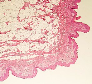
Histopathology, showing thin rim of epidermis, and dermal lipocytes without capsule.

.jpg)
Microscopy/Histopathology report
For lipomas: Absence of signs of malignancy.
 ((Tissue composed of univacuolar fat cells and delicate and inconspicuous fibrous septa.)) No evidence of malignancy. |
See also: General notes on reporting
Notes
- ↑ For a full list of contributors, see article history. Creators of images are attributed at the image description pages, seen by clicking on the images. See Patholines:Authorship for details.
Main page
References
- ↑ Monica Dahlgren, Janne Malina, Anna Måsbäck, Otto Ljungberg (1997-02-13). Lilla utskärningen.
- ↑ 2.0 2.1 Andreas F Mavrogenis, Panayiotis J Papagelopoulos (2013-02-01). Soft Tissues: Well-differentiated liposarcoma. Atlas of Genetics and Cytogenetics in Oncology and Haematology.
- ↑ Pathology Department at NU Hospital Group, Sweden, 2019-2020.
- ↑ . Lipoma. Gross Pathology Manual - By The University of Chicago Department of Pathology. Retrieved on 2020-08-26.
- ↑ . Lipoma Variant: Fibrolipoma. Stanford University School of Medicine. Retrieved on 2020-02-10.
- ↑ Vijay Shankar. Soft tissue - Adipose tissue tumors - Lipoma and variants - Angiolipoma. Pathology Outlines. Topic Completed: 1 August 2012. Minor changes: 20 March 2019
- ↑ Michael R. Clay, M.D.. Liposarcoma. PathologyOutlines. Topic Completed: 1 November 2017. Minor changes: 11 May 2021
- ↑ Michael R. Clay. Soft tissue - Adipose tissue - Myxoid liposarcoma. PathologyOutlines. Topic Completed: 1 January 2018. Revised: 20 March 2019
Image sources