Difference between revisions of "Liver"
Jump to navigation
Jump to search
(Not that relevant for a pathologist) |
(Expanded) |
||
| Line 6: | Line 6: | ||
*'''[[Autopsy]]''' | *'''[[Autopsy]]''' | ||
*Liver biopsy | *Liver biopsy | ||
| + | |||
| + | {{Fixation - standard}} | ||
| + | Non–formalin-fixed tissue may be needed for tests such as microbiological analysis or copper quantification studies.<ref name="BoydCain2020">{{cite journal|last1=Boyd|first1=Alexander|last2=Cain|first2=Owen|last3=Chauhan|first3=Abhishek|last4=Webb|first4=Gwilym James|title=Medical liver biopsy: background, indications, procedure and histopathology|journal=Frontline Gastroenterology|volume=11|issue=1|year=2020|pages=40–47|issn=2041-4137|doi=10.1136/flgastro-2018-101139}}</ref> | ||
==Gross processing in autopsy== | ==Gross processing in autopsy== | ||
Revision as of 04:02, 20 December 2019
Author:
Mikael Häggström [note 1]
Contents
Tissue sampling
- Autopsy
- Liver biopsy
Fixation
Generally 10% neutral buffered formalin. Non–formalin-fixed tissue may be needed for tests such as microbiological analysis or copper quantification studies.[1]
Gross processing in autopsy
Make consecutive liver slices, such as in the sagittal or coronal plane.
Basic gross examination
- Inspect the color and texture of the surfaces, including external and cut surfaces. Potential pathologies:

Diffuse areas of pallor in cirrhosis, see Cirrhosis
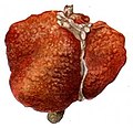
Pale macronodules of cirrhosis, see Cirrhosis

Nutmeg texture of congestive hepatopathy

Liver metastases

.jpg)
.jpg)
.jpg)
- Look for any focal change in the liver volume, mainly any tumor.
- Determine liver weight.
Gross report
- Weight
- Color and texture of cut surfaces
- Any focal change
Notes
- ↑ For a full list of contributors, see article history. Creators of images are attributed at the image description pages, seen by clicking on the images. See Patholines:Authorship for details.
Main page
References
Image sources