Difference between revisions of "Liver tumor"
Jump to navigation
Jump to search

(Started) |
(→Microscopic evaluation: +Images) |
||
| Line 18: | Line 18: | ||
==Microscopic evaluation== | ==Microscopic evaluation== | ||
[[File:Liver tumor types in adults by relative incidence.png|thumb|Liver tumor types by relative incidence in adults in the United States.<ref name=Sternberg>[https://basicmedicalkey.com/masses-of-the-liver-2/ Table 37.2] in: {{cite book | last=Sternberg | first=Stephen | title=Sternberg's diagnostic surgical pathology | publisher=LWW | publication-place=Place of publication not identified | year=2012 | isbn=978-1-4511-5289-0 | oclc=953861627 | ref=harv}}</ref>]] | [[File:Liver tumor types in adults by relative incidence.png|thumb|Liver tumor types by relative incidence in adults in the United States.<ref name=Sternberg>[https://basicmedicalkey.com/masses-of-the-liver-2/ Table 37.2] in: {{cite book | last=Sternberg | first=Stephen | title=Sternberg's diagnostic surgical pathology | publisher=LWW | publication-place=Place of publication not identified | year=2012 | isbn=978-1-4511-5289-0 | oclc=953861627 | ref=harv}}</ref>]] | ||
| − | Hepatocellular carcinoma is the most common diagnosis for liver tumors,<ref name=Sternberg/> so start by looking for characteristics thereof. | + | Hepatocellular carcinoma is the most common diagnosis for liver tumors,<ref name=Sternberg/> so start by looking for characteristics thereof: |
| + | |||
| + | '''Hepatocellular carcinoma:''' | ||
| + | <gallery> | ||
| + | File:Hepatocellular carcinoma (13896532458).jpg|Needle biopsy. | ||
| + | File:Hepatocellular carcinoma (13896504027).jpg|Needle biopsy. | ||
| + | File:Hepatocellular carcinoma (14083110265).jpg|Needle biopsy. | ||
| + | File:Hepatocellular Carcinoma, FNA (2330701083).jpg|FNA. | ||
| + | File:Hepatocellular Carcinoma, FNA Clot Section (2330699911).jpg|FNA clot section. | ||
| + | File:Hepatocellular Carcinoma, FNA Clot Section (2331528214).jpg|FNA clot section. | ||
| + | </gallery> | ||
{{Further|Evaluation of suspected malignancies}} | {{Further|Evaluation of suspected malignancies}} | ||
{{Bottom}} | {{Bottom}} | ||
Revision as of 11:31, 24 February 2020
Author:
Mikael Häggström [note 1]
Contents
Tissue sampling
- Liver biopsy
- Autopsy: Further information: Autopsy
Gross examination
Note the following:[1]
- Whether the tumor is sell demarcated from surrounding tissue
- Whether there is visible infiltration or invasion into surrounding tissue
- Any necrosis or bleeding

Hepatocellular carcinoma

Microscopic evaluation

Liver tumor types by relative incidence in adults in the United States.[2]
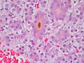
Hepatocellular carcinoma is the most common diagnosis for liver tumors,[2] so start by looking for characteristics thereof:
Hepatocellular carcinoma:

Needle biopsy.

Needle biopsy.

Needle biopsy.

FNA.

FNA clot section.
FNA clot section.
.jpg)
.jpg)
.jpg)
.jpg)
.jpg)
.jpg)
Further information: Evaluation of suspected malignancies
Notes
- ↑ For a full list of contributors, see article history. Creators of images are attributed at the image description pages, seen by clicking on the images. See Patholines:Authorship for details.
Main page
References
- ↑ . General oncology. Amboss. Retrieved on 2020-01-29.
- ↑ 2.0 2.1 Table 37.2 in: Sternberg, Stephen (2012). Sternberg's diagnostic surgical pathology . Place of publication not identified: LWW. ISBN 978-1-4511-5289-0. OCLC 953861627.
Image sources