Difference between revisions of "Lung autopsy"
Jump to navigation
Jump to search

(→Gross evaluation: +Normal weight) |
(→Microscopic evaluation: Better) |
||
| Line 36: | Line 36: | ||
==Microscopic evaluation== | ==Microscopic evaluation== | ||
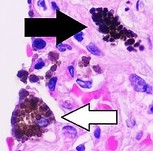
| − | [[File:Histopathology of | + | [[File:Histopathology of lung siderophages, annotated.jpg|thumb|220px|'''Siderophages'''{{Siderophage note 1}} (one indicated by white arrows), indicating chronic left heart failure (but may also be seen after pulmonary hemorrhage). Also pulmonary '''congestion''' and '''lymphocytes''', consistent with acute or chronic heart failure.]] |
Look for the most common pathologic lung findings:<ref name="TiwanaNibhoria2014">'''India''': {{cite journal|last1=Tiwana|first1=Kanwardeep Kaur|last2=Nibhoria|first2=Sarita|last3=Gupta|first3=Manvi|last4=Yadav|first4=Ashish|title=Histopathological Spectrum in Lung Autopsies- A 50 Case Study|journal=Indian Journal of Forensic Medicine & Toxicology|volume=8|issue=2|year=2014|pages=172|issn=0973-9122|doi=10.5958/0973-9130.2014.00709.9}}</ref><ref>'''United States''': {{cite web|url=https://washingtonforensicsservices.com/pulmonary-lung-conditions-found-at-autopsy/|title=Pulmonary Lung Conditions Found at Autopsy|author=Dr. Stanley Adams|website=Washington Forensic Services|accessdate=2019-12-20}}</ref> | Look for the most common pathologic lung findings:<ref name="TiwanaNibhoria2014">'''India''': {{cite journal|last1=Tiwana|first1=Kanwardeep Kaur|last2=Nibhoria|first2=Sarita|last3=Gupta|first3=Manvi|last4=Yadav|first4=Ashish|title=Histopathological Spectrum in Lung Autopsies- A 50 Case Study|journal=Indian Journal of Forensic Medicine & Toxicology|volume=8|issue=2|year=2014|pages=172|issn=0973-9122|doi=10.5958/0973-9130.2014.00709.9}}</ref><ref>'''United States''': {{cite web|url=https://washingtonforensicsservices.com/pulmonary-lung-conditions-found-at-autopsy/|title=Pulmonary Lung Conditions Found at Autopsy|author=Dr. Stanley Adams|website=Washington Forensic Services|accessdate=2019-12-20}}</ref> | ||
*'''[[Alveolar fluid]]'''. {{further|Alveolar fluid|linebreak=no}} | *'''[[Alveolar fluid]]'''. {{further|Alveolar fluid|linebreak=no}} | ||
Revision as of 19:12, 18 December 2020
Author:
Mikael Häggström [note 1]
Autopsy of the lungs, not including larger pulmonary vessels (instead summarized at Autopsy - Other thorax).
Contents
Basic autopsy cutting
In non-forensic Autopsy:
- The lungs may be cut after removing the heart through cutting through the major vessels close to it, or by removing each lung by cuts by each lung hilum.
- Dissect the pulmonary arterial system, from the pulmonary trunk and including at least segmental arteries.
- Dissect the bronchial tree, at least to segmental bronchi. Check for obstructions.
- Weigh each lung (possibly first if having cut each lung at the hilus).
- Make some additional sections through the lung parenchyma. Squeeze at each side to detect any pus and edema.[1]
- For context, see Autopsy
Gross evaluation

- A spongy consistency, and watery and frothy liquid being pressed from the parenchyma, indicates simple edema.[2]
- A spongy consistency and reddish (blood-stained) fluid being pressed from the parenchyma, indicates acute congestion.[2]
- A brownish or dark reddish color of the fluid pressed from the parenchyma indicates chronic congestion, and may not have a spongy consistency.[2]
Normal weight:
| Left | Right | |
|---|---|---|
| Men[3] | 112-675g | 155-720g |
| Women[4] | 105-515g | 101-589g |
Fixation
Generally 10% neutral buffered formalin.
See also: General notes on fixation
Microscopic evaluation

Siderophages[notes 1] (one indicated by white arrows), indicating chronic left heart failure (but may also be seen after pulmonary hemorrhage). Also pulmonary congestion and lymphocytes, consistent with acute or chronic heart failure.
Look for the most common pathologic lung findings:[5][6]
- Alveolar fluid. Further information: Alveolar fluid
- Vascular congestion, which can usually be seen easiest in the alveolar walls. It indicates left sided heart failure, especially when seen together with alveolar fluid.
- Inflammatory cells, where a mild to moderate lymphocytic infiltrate is consistent with with heart failure, while neutrophils indicate pneumonia.
- Mycobacteria in regions of the world with substantial prevalence
- Carcinoma Further information: Lung tumor
- Aspiration: Other foreign contents in airways. Further information: Aspiration in autopsy

Edema

Bronchopneumonia, with neutrophils filling a bronchiole.

Carcinoma (in this case bronchioloalveolar cell adenocarcinoma) Further information: Lung tumor


Main diagnoses
- Left sided heart failure:

If respiratory epithelial shedding is seen, look for vascular leakage, mucus hypersecretion and/or widespread airway narrowing, together indicating asthma death.[8] Otherwise, it is a frequent postmortem change.
Additional potential findings are mentioned in the general Lungs article.
Reporting
Report findings and if they are consistent with already known diagnoses.
Example:
 |
Further information: Autopsy
Notes
- ↑ For a full list of contributors, see article history. Creators of images are attributed at the image description pages, seen by clicking on the images. See Patholines:Authorship for details.
Main page
References
- ↑ Burton, Julian L.; Rutty, Guy N. (2010). The Hospital Autopsy A Manual of Fundamental Autopsy Practice (3rd ed.). Oxford University Press. ISBN 978-0340965146.
- ↑ 2.0 2.1 2.2 page 62 in: J. Martin Beattie (2014). Post-Mortem Methods . Cambridge University Press. ISBN 9781107418004.
- ↑ Molina, D. Kimberley; DiMaio, Vincent J.M. (2012). "Normal Organ Weights in Men ". The American Journal of Forensic Medicine and Pathology 33 (4): 368–372. doi:. ISSN 0195-7910.
- ↑ Molina, D. Kimberley; DiMaio, Vincent J. M. (2015). "Normal Organ Weights in Women ". The American Journal of Forensic Medicine and Pathology 36 (3): 182–187. doi:. ISSN 0195-7910.
- ↑ India: Tiwana, Kanwardeep Kaur; Nibhoria, Sarita; Gupta, Manvi; Yadav, Ashish (2014). "Histopathological Spectrum in Lung Autopsies- A 50 Case Study ". Indian Journal of Forensic Medicine & Toxicology 8 (2): 172. doi:. ISSN 0973-9122.
- ↑ United States: Dr. Stanley Adams. Pulmonary Lung Conditions Found at Autopsy. Washington Forensic Services. Retrieved on 2019-12-20.
- ↑ 7.0 7.1 . Congestion. Humpath (2005-12-19).
- ↑ Madea, B (2014). Handbook of forensic medicine . Hoboken, N.J: Wiley-Blackwell. ISBN 978-1-118-57062-3. OCLC 872114659.
Image sources