Lung autopsy
Author:
Mikael Häggström [note 1]
Autopsy of the lungs, not including larger pulmonary vessels (instead summarized at Autopsy - Other thorax).
Contents
Basic autopsy cutting
In non-forensic Autopsy:
- The lungs may be cut after removing the heart through cutting through the major vessels close to it, or by removing each lung by cuts by each lung hilum.
- Dissect the pulmonary arterial system, from the pulmonary trunk and including at least segmental arteries.
- Dissect the bronchial tree, at least to segmental bronchi. Check for obstructions.
- Weigh each lung (possibly first if having cut each lung at the hilus).
- Make some additional sections through the lung parenchyma. Squeeze at each side to detect any pus and edema.[1]
- For context, see Autopsy
Gross evaluation

- A spongy consistency, and watery and frothy liquid being pressed from the parenchyma, indicates simple edema.[2]
- A spongy consistency and reddish (blood-stained) fluid being pressed from the parenchyma, indicates acute congestion.[2]
- A brownish or dark reddish color of the fluid pressed from the parenchyma indicates chronic congestion, and may not have a spongy consistency.[2]
Normal weight:
| Left | Right | |
|---|---|---|
| Men[3] | 112-675g | 155-720g |
| Women[4] | 105-515g | 101-589g |
Fixation
Generally 10% neutral buffered formalin.
See also: General notes on fixation
Microscopic evaluation
Look for the most common pathologic lung findings:[5][6]
- Alveolar fluid. Further information: Alveolar fluid
- Vascular congestion, which can usually be seen easiest in the alveolar walls. It indicates left sided heart failure, especially when seen together with alveolar fluid. Further information: Chronic pulmonary congestion
- Inflammatory cells, where a mild to moderate lymphocytic infiltrate is consistent with with heart failure, while neutrophils indicate pneumonia. pigmented macrophages of the lung may indicate chronic heart failure.
- Mycobacteria in regions of the world with substantial prevalence
- Carcinoma Further information: Lung tumor
- Aspiration: Other foreign contents in airways. Further information: Aspiration in autopsy
- Embolism of pulmonary arteries.

Edema

Bronchopneumonia, with neutrophils filling a bronchiole.

Carcinoma (in this case bronchioloalveolar cell adenocarcinoma) Further information: Lung tumor

hyaline membranes, suggesting diffuse alveolar damage.
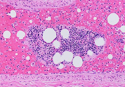
A pulmonary artery with fat embolism (seen as multiple empty globular spaces on this H&E stain since its processing dissolves fat). There is a bone marrow fragment in the middle, and multiple single hematopoietic cells in the blood, being evidence of fracture as the source of the embolism.


Main diagnoses
- Left sided heart failure:
- Acute congestion manifests as alveolar capillaries being engorged with blood, as well as associated alveolar septal edema and/or focal intra-alveolar hemorrhage.[7]
- Chronic pulmonary congestion manifests as thickened and fibrotic septa, and alveolar spaces containing numerous pigmented lung macrophages.[7]

Additional potential findings are mentioned in the general Lungs article.
Reporting
Report findings and if they are consistent with already known diagnoses.
Example:
 |
Further information: Autopsy
Notes
- ↑ For a full list of contributors, see article history. Creators of images are attributed at the image description pages, seen by clicking on the images. See Patholines:Authorship for details.
Main page
References
- ↑ Burton, Julian L.; Rutty, Guy N. (2010). The Hospital Autopsy A Manual of Fundamental Autopsy Practice (3rd ed.). Oxford University Press. ISBN 978-0340965146.
- ↑ 2.0 2.1 2.2 page 62 in: J. Martin Beattie (2014). Post-Mortem Methods . Cambridge University Press. ISBN 9781107418004.
- ↑ Standard reference range: Molina, D. Kimberley; DiMaio, Vincent J.M. (2012). "Normal Organ Weights in Men ". The American Journal of Forensic Medicine and Pathology 33 (4): 368–372. doi:. ISSN 0195-7910.
- ↑ Standard reference range: Molina, D. Kimberley; DiMaio, Vincent J. M. (2015). "Normal Organ Weights in Women ". The American Journal of Forensic Medicine and Pathology 36 (3): 182–187. doi:. ISSN 0195-7910.
- ↑ India: Tiwana, Kanwardeep Kaur; Nibhoria, Sarita; Gupta, Manvi; Yadav, Ashish (2014). "Histopathological Spectrum in Lung Autopsies- A 50 Case Study ". Indian Journal of Forensic Medicine & Toxicology 8 (2): 172. doi:. ISSN 0973-9122.
- ↑ United States: Dr. Stanley Adams. Pulmonary Lung Conditions Found at Autopsy. Washington Forensic Services. Retrieved on 2019-12-20.
- ↑ 7.0 7.1 . Congestion. Humpath (2005-12-19).
- ↑ Madea, B (2014). Handbook of forensic medicine . Hoboken, N.J: Wiley-Blackwell. ISBN 978-1-118-57062-3. OCLC 872114659.
Image sources