Difference between revisions of "Lung tumor"
Jump to navigation
Jump to search
(Expanded) |
(→Gross processing: Vascular) |
||
| Line 18: | Line 18: | ||
*Measure '''tumor size''' as a maximum diameter {{Moderate-begin}}or 3 dimensions{{Moderate-end}} | *Measure '''tumor size''' as a maximum diameter {{Moderate-begin}}or 3 dimensions{{Moderate-end}} | ||
*Determine '''location''': Which lobe if applicable, and if it is peripheral, central or hilar. | *Determine '''location''': Which lobe if applicable, and if it is peripheral, central or hilar. | ||
| − | *'''Margin''' length to pleura and hilum/surgical margin. | + | *'''Margin''' length to pleura and hilum/surgical margin. {{Moderate-begin}}In wedge resections, also measure the distance to the bronchial/vascular margin.{{Moderate-end}} |
*Any '''involvement''' of major bronchi or blood vessels. | *Any '''involvement''' of major bronchi or blood vessels. | ||
*Any abnormal '''lymph nodes''', including hilar ones if available. | *Any abnormal '''lymph nodes''', including hilar ones if available. | ||
Revision as of 09:59, 17 March 2021
Author:
Mikael Häggström [note 1]
Contents
Comprehensiveness
On this resource, the following formatting is used for comprehensiveness:
- Minimal depth
- (Moderate depth)
- ((Comprehensive))
Presentations
- Bronchial lavage
- Lung needle biopsy
- Lung wedge resection or lobectomy
- Lung autopsy
Gross processing
In tumor resection:[1]
- Measure the specimen in 3 dimensions.
- Describe any included pleural surface, including color, and any presence of granularity, adhesions, retraction, or tumor.
- Serially section the specimen. Describe the cut surface, including color and consistency, and any focal lesions.
- Measure tumor size as a maximum diameter (or 3 dimensions)
- Determine location: Which lobe if applicable, and if it is peripheral, central or hilar.
- Margin length to pleura and hilum/surgical margin. (In wedge resections, also measure the distance to the bronchial/vascular margin.)
- Any involvement of major bronchi or blood vessels.
- Any abnormal lymph nodes, including hilar ones if available.
Photograph all tumors.
Microscopic evaluation
Medical imaging provides a major clue as to whether a lung tumor is benign or malignant, where lesions smaller than 2 cm are likely to be benign, whereas lesions larger than 2 cm are malignant (that is, lung cancer) in 85% of cases.[2]
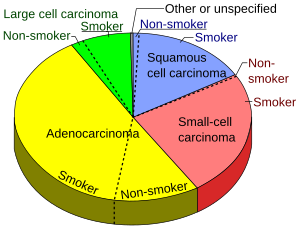
Subsequently distribution of benign tumors and lung cancers, respectively, are as follows:[2]
Benign lung tumors:
- Hamartomas - 76%
- Benign fibrous mesothelioma/solitary fibrous tumor (SFT) - 12.3%
- Inflammatory pseudotumor (IPT) - 5.4%
- Lipoma - 1.5%
- Leiomyoma - 1.5%
- Other - 3.3%
Gallery of lung cancers
Squamous-cell carcinoma of the lung. Typical squamous-cell carcinoma cells are large with abundant eosinophilic cytoplasm and large, often vesicular, nuclei.[3]
Notes
- ↑ For a full list of contributors, see article history. Creators of images are attributed at the image description pages, seen by clicking on the images. See Patholines:Authorship for details.
Main page
References
- ↑ . Pulmonary pathology grossing guidelines. Retrieved on 2021-03-17.
- ↑ 2.0 2.1 Alain C. Borczuk (2008). "Benign Tumors and Tumorlike Conditions of the Lung ". Archives of Pathology & Laboratory Medicine 132 (7). Archived from the original. .
- ↑ Dr Nicholas Turnbull, A/Prof Patrick Emanual (2014-05-03). Squamous cell carcinoma pathology. DermNetz.
Image sources


