Difference between revisions of "Lung tumor"
(→Gallery of lung cancers: Described) |
(→Gallery of lung cancers: Linked away) |
||
| Line 29: | Line 29: | ||
===Gallery of lung cancers=== | ===Gallery of lung cancers=== | ||
<gallery mode=packed heights=190> | <gallery mode=packed heights=190> | ||
| − | File:Lung adenocarcinoma with lepidic growth - low magnification.jpg|'''Lung adenocarcinoma''', with lepidic pattern shown, wherein tumors cells cover alveolar walls. | + | File:Lung adenocarcinoma with lepidic growth - low magnification.jpg|'''[[Lung adenocarcinoma]]''', with lepidic pattern shown, wherein tumors cells cover alveolar walls. |
| + | File:Histopathology of lung adenocarcinoma with solid pattern.jpg|'''[[Lung adenocarcinoma]]''', with solid pattern. | ||
File:Large cell carcinoma of the lung .jpg|'''Large cell carcinoma''' of the lung: neoplastic cells with abundant pale eosinophilic cytoplasm | File:Large cell carcinoma of the lung .jpg|'''Large cell carcinoma''' of the lung: neoplastic cells with abundant pale eosinophilic cytoplasm | ||
File:Histopathology of squamous-cell carcinoma of the lung.jpg|'''[[Squamous-cell carcinoma of the lung]]'''. Typical squamous-cell carcinoma cells are large with abundant eosinophilic cytoplasm and large, often vesicular, nuclei.<ref>{{cite web|url=https://dermnetnz.org/topics/squamous-cell-carcinoma-pathology/|title=Squamous cell carcinoma pathology|website=DermNetz|author=Dr Nicholas Turnbull, A/Prof Patrick Emanual|date=2014-05-03}}</ref> | File:Histopathology of squamous-cell carcinoma of the lung.jpg|'''[[Squamous-cell carcinoma of the lung]]'''. Typical squamous-cell carcinoma cells are large with abundant eosinophilic cytoplasm and large, often vesicular, nuclei.<ref>{{cite web|url=https://dermnetnz.org/topics/squamous-cell-carcinoma-pathology/|title=Squamous cell carcinoma pathology|website=DermNetz|author=Dr Nicholas Turnbull, A/Prof Patrick Emanual|date=2014-05-03}}</ref> | ||
</gallery> | </gallery> | ||
| − | |||
| − | |||
{{Bottom}} | {{Bottom}} | ||
Revision as of 16:15, 3 July 2021
Author:
Mikael Häggström [note 1]
Contents
Comprehensiveness
On this resource, the following formatting is used for comprehensiveness:
- Minimal depth
- (Moderate depth)
- ((Comprehensive))
Presentations
- Bronchial lavage
- Lung needle biopsy
- Lung wedge resection or lobectomy
- Lung autopsy
Gross processing
As per presentation above.
Microscopic evaluation
Medical imaging provides a major clue as to whether a lung tumor is benign or malignant, where lesions smaller than 2 cm are likely to be benign, whereas lesions larger than 2 cm are malignant (that is, lung cancer) in 85% of cases.[1]
Subsequently distribution of benign tumors and lung cancers, respectively, are as follows:[1]
Benign lung tumors:
- Hamartomas - 76%
- Benign fibrous mesothelioma/solitary fibrous tumor (SFT) - 12.3%
- Inflammatory pseudotumor (IPT) - 5.4%
- Lipoma - 1.5%
- Leiomyoma - 1.5%
- Other - 3.3%
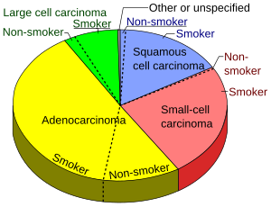
Gallery of lung cancers
Lung adenocarcinoma, with lepidic pattern shown, wherein tumors cells cover alveolar walls.
Lung adenocarcinoma, with solid pattern.
Squamous-cell carcinoma of the lung. Typical squamous-cell carcinoma cells are large with abundant eosinophilic cytoplasm and large, often vesicular, nuclei.[2]
Notes
- ↑ For a full list of contributors, see article history. Creators of images are attributed at the image description pages, seen by clicking on the images. See Patholines:Authorship for details.
Main page
References
- ↑ 1.0 1.1 Alain C. Borczuk (2008). "Benign Tumors and Tumorlike Conditions of the Lung ". Archives of Pathology & Laboratory Medicine 132 (7). Archived from the original. .
- ↑ Dr Nicholas Turnbull, A/Prof Patrick Emanual (2014-05-03). Squamous cell carcinoma pathology. DermNetz.
Image sources


