Difference between revisions of "Myocardial fibrosis"
(→Microscopic evaluation: HE stain) |
(→Microscopic evaluation: +Fat deposition) |
||
| Line 16: | Line 16: | ||
File:Subepicardial fibrosis.jpg|'''Subepicardial fibrosis''' (epicardium at top). | File:Subepicardial fibrosis.jpg|'''Subepicardial fibrosis''' (epicardium at top). | ||
File:Histopathology of dense fibrous scar replacing myocyte loss in myocardial infarction.jpg|'''Replacement fibrosis''' in myocardial infarction, typically being boundless, dense and not conforming to the other types. | File:Histopathology of dense fibrous scar replacing myocyte loss in myocardial infarction.jpg|'''Replacement fibrosis''' in myocardial infarction, typically being boundless, dense and not conforming to the other types. | ||
| + | File:Histopathology of fat deposition in myocardial infarction scar.jpg|Fat deposition further indicates '''replacement fibrosis''' of a previous myocardial infarction.<ref name="ChenitiSridi2019">{{cite journal|last1=Cheniti|first1=Ghassen|last2=Sridi|first2=Soumaya|last3=Sacher|first3=Frederic|last4=Chaumeil|first4=Arnaud|last5=Pillois|first5=Xavier|last6=Takigawa|first6=Masateru|last7=Frontera|first7=Antonio|last8=Vlachos|first8=Konstantinos|last9=Martin|first9=Claire A.|last10=Teijeira|first10=Elvis|last11=Kitamura|first11=Takeshi|last12=Lam|first12=Anna|last13=Bourier|first13=Felix|last14=Puyo|first14=Stephane|last15=Duchateau|first15=Josselin|last16=Denis|first16=Arnaud|last17=Pambrun|first17=Thomas|last18=Chauvel|first18=Remi|last19=Derval|first19=Nicolas|last20=Laurent|first20=François|last21=Montaudon|first21=Michel|last22=Hocini|first22=Meleze|last23=Haissaguerre|first23=Michel|last24=Jais|first24=Pierre|last25=Cochet|first25=Hubert|title=Post–Myocardial Infarction Scar With Fat Deposition Shows Specific Electrophysiological Properties and Worse Outcome After Ventricular Tachycardia Ablation|journal=Journal of the American Heart Association|volume=8|issue=15|year=2019|issn=2047-9980|doi=10.1161/JAHA.119.012482}}</ref> | ||
</gallery> | </gallery> | ||
Revision as of 17:33, 21 May 2021
Author:
Mikael Häggström [note 1]
Contents
Presentations
Microscopic evaluation
In case of myocardial fibrosis, attempt to distinguish between the following:
- Interstitial fibrosis, which is unspecific,[1] (and may simply reported as such).[notes 1]
- Subepicardial fibrosis, also unspecific (and may simply be reported as such).[notes 2]
- Replacement fibrosis, which indicates an older infarction.[1]

Healthy myocardium versus interstitial fibrosis in dilated cardiomyopathy. Alcian blue stain. The fibrosis is either evenly distributed between myocytes or follows anatomic structures such as blood vessels.

Interstitial fibrosis of chronic ischemic heart disease, H&E stain, with associated relatively well organized myocardial bundles
Subepicardial fibrosis (epicardium at top).

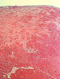
Replacement fibrosis in myocardial infarction, typically being boundless, dense and not conforming to the other types.

Fat deposition further indicates replacement fibrosis of a previous myocardial infarction.[2]


Reporting
Example (if uncertain whether there is replacement fibrosis or merely subepicardial fibrosis):
 |
Notes
- ↑ Interstitial fibrosis has been described in congestive heart failure, hypertension, and normal aging. (Chute, 2019)
- ↑ Subepicardial fibrosis is associated with non-infarction diagnoses such as myocarditis and non-ischemic cardiomyopathy.
- Gräni, Christoph; Eichhorn, Christian; Bière, Loïc; Kaneko, Kyoichi; Murthy, Venkatesh L.; Agarwal, Vikram; Aghayev, Ayaz; Steigner, Michael; et al. (2019). "Comparison of myocardial fibrosis quantification methods by cardiovascular magnetic resonance imaging for risk stratification of patients with suspected myocarditis ". Journal of Cardiovascular Magnetic Resonance 21 (1). doi:. ISSN 1532-429X.
- Bhaskaran, Ashwin; Tung, Roderick; Stevenson, William G.; Kumar, Saurabh (2019). "Catheter Ablation of VT in Non-Ischaemic Cardiomyopathies: Endocardial, Epicardial and Intramural Approaches ". Heart, Lung and Circulation 28 (1): 84–101. doi:. ISSN 14439506.
- ↑ For a full list of contributors, see article history. Creators of images are attributed at the image description pages, seen by clicking on the images. See Patholines:Authorship for details.
Main page
References
- ↑ 1.0 1.1 Chute, Michael; Aujla, Preetinder; Jana, Sayantan; Kassiri, Zamaneh (2019). "The Non-Fibrillar Side of Fibrosis: Contribution of the Basement Membrane, Proteoglycans, and Glycoproteins to Myocardial Fibrosis ". Journal of Cardiovascular Development and Disease 6 (4): 35. doi:. ISSN 2308-3425.
- ↑ Cheniti, Ghassen; Sridi, Soumaya; Sacher, Frederic; Chaumeil, Arnaud; Pillois, Xavier; Takigawa, Masateru; Frontera, Antonio; Vlachos, Konstantinos; et al. (2019). "Post–Myocardial Infarction Scar With Fat Deposition Shows Specific Electrophysiological Properties and Worse Outcome After Ventricular Tachycardia Ablation ". Journal of the American Heart Association 8 (15). doi:. ISSN 2047-9980.
Image sources