Difference between revisions of "Nasal cavity and paranasal sinuses"
Jump to navigation
Jump to search

(Replaced) Tags: Mobile web edit, Mobile edit |
m (→Nasal or sinonasal polyps: Bolded) |
||
| Line 14: | Line 14: | ||
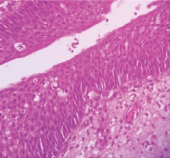
File:Histopathology of nasal squamous papilloma.jpg|'''Squamous papilloma''', with acanthosis and hyperkerratosis. | File:Histopathology of nasal squamous papilloma.jpg|'''Squamous papilloma''', with acanthosis and hyperkerratosis. | ||
</gallery> | </gallery> | ||
| + | In case of '''inflammation''', confirm that it is mixed and that lymphocytes are not atypical (otherwise, consult hematopathology, particularly whether it could be T cell lymphoma, nasal type. | ||
{{Bottom}} | {{Bottom}} | ||
Revision as of 19:23, 26 October 2022
Author:
Mikael Häggström [note 1]
Nasal or sinonasal polyps
Look for signs of malignancy. Further information: Evaluation of suspected malignancies

Benign nasal/sinonasal polyp (not otherwise specified), consisting of hyperplastic edematous connective tissue with some seromucous glands and inflammation (mostly neutrophils and eosinophils), surrounded by respiratory or squamous epithelium.[1] It can be termed inflammatory nasal/sinonasal polyp when inflammation is more pronounced.
Main differential diagnoses:
Inverted papilloma, wherein the surface epithelial cells grow downward into the underlying supportive tissue.

Squamous papilloma, with acanthosis and hyperkerratosis.


In case of inflammation, confirm that it is mixed and that lymphocytes are not atypical (otherwise, consult hematopathology, particularly whether it could be T cell lymphoma, nasal type.
Notes
- ↑ For a full list of contributors, see article history. Creators of images are attributed at the image description pages, seen by clicking on the images. See Patholines:Authorship for details.
Main page
References
- ↑ Michaels, Leslie (2012-12-06) (in en). Ear, Nose and Throat Histopathology . Springer Science & Business Media. p. 168. ISBN 9781447133322.
Image sources