Ovary
Revision as of 10:02, 24 November 2020 by Mikael Häggström (talk | contribs) (→Gross processing: No lesions)
Author:
Mikael Häggström [note 1]
Contents
Fixation
Generally 10% neutral buffered formalin.
Presentations
Gross processing

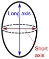
"Long" and "short" axis.[1]
The ovary is cut in the longitudinal plane (through the "long axis").
Gross report
Template:
| (A. Labeled - __. The specimen is received in formalin and consists of) an ovary measuring ___. The ovarian capsule is tan-pink and smooth. Cut sections reveal solid, white and whorled parenchyma, and no gross lesions. Representative sections are submitted for microscopic examination in __ cassettes. |
See also
Notes
- ↑ For a full list of contributors, see article history. Creators of images are attributed at the image description pages, seen by clicking on the images. See Patholines:Authorship for details.
Main page
References
Image sources