Paratubal cyst
Author:
Mikael Häggström [note 1]
Paratubal cysts are also called paraovarian cysts, depending on location.[1]
Contents
Comprehensiveness
On this resource, the following formatting is used for comprehensiveness:
- Minimal depth
- (Moderate depth)
- ((Comprehensive))
Gross processing
Paratubal cysts are generally incidentally found on grossing of fallopian tubes, and in such cases they may be ignored, as they are essentially always benign.[2] ( or included in representative sections). Even more insignificant are sessile smaller cyst-like along the fallopian tube, which are serosal inclusion cysts.
Microscopic examination
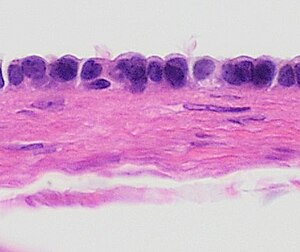
Paratubal cysts are generally lined by simple cuboidal epithelium as shown. However, they may have fallopian tubal epithelium or focal papillary projections.[1]
Notes
- ↑ For a full list of contributors, see article history. Creators of images are attributed at the image description pages, seen by clicking on the images. See Patholines:Authorship for details.
Main page
References
- ↑ 1.0 1.1 Nicole Riddle, Jamie Shutter. Fallopian tubes & broad ligament, Broad ligament, Paratubal cysts. Pathology Outlines. Topic Completed: 1 July 2013. Minor changes: 30 December 2020
- ↑ Shin, You-Jung; Kim, Ji-Young; Lee, Hee Jin; Park, Jeong-Yeol; Nam, Joo-Hyun (2011). "Paratubal serous borderline tumor ". Journal of Gynecologic Oncology 22 (4): 295. doi:. ISSN 2005-0380.
Image sources

