Difference between revisions of "Smooth muscle tumor"
(→Microscopic examination: +Criteria) |
(→Microscopic examination: +Pleomorphism) |
||
| Line 31: | Line 31: | ||
File:Histopathology of uterine leiomyoma.jpg|'''Leiomyoma'''s typically show smooth muscle in a whorled (fascicular) pattern<ref>{{cite web|url=http://www.pathologyoutlines.com/topic/uterusleiomyoma.html|title=Uterus - Stromal tumors - Leiomyoma|author=Mohamed Mokhtar Desouki|website=Pathology Outlines}} Topic Completed: 1 August 2011. Revised: 15 December 2019</ref> | File:Histopathology of uterine leiomyoma.jpg|'''Leiomyoma'''s typically show smooth muscle in a whorled (fascicular) pattern<ref>{{cite web|url=http://www.pathologyoutlines.com/topic/uterusleiomyoma.html|title=Uterus - Stromal tumors - Leiomyoma|author=Mohamed Mokhtar Desouki|website=Pathology Outlines}} Topic Completed: 1 August 2011. Revised: 15 December 2019</ref> | ||
File:Histopathology of a leiomyoma in a postmenopausal uterus, intermediate magnification.jpg|'''Leiomyoma''', with areas where the cellularity is relatively lower (left) and higher (right). | File:Histopathology of a leiomyoma in a postmenopausal uterus, intermediate magnification.jpg|'''Leiomyoma''', with areas where the cellularity is relatively lower (left) and higher (right). | ||
| + | File:Histopathology of leiomyoma with nuclear pleomorphism.jpg|thumb|'''Leiomyoma''' with nuclear pleomorphism, yet within benign limits. | ||
File:Epithelioid Leiomyoma, Uterus (3) (2556674506).jpg|Epithelioid '''leiomyoma''' | File:Epithelioid Leiomyoma, Uterus (3) (2556674506).jpg|Epithelioid '''leiomyoma''' | ||
File:Leiomyoma of Uterus with Palisading Pattern (5710417630).jpg|'''Leiomyoma''' with palisading pattern | File:Leiomyoma of Uterus with Palisading Pattern (5710417630).jpg|'''Leiomyoma''' with palisading pattern | ||
Revision as of 17:37, 2 May 2021
Author:
Mikael Häggström [note 1]
Contents
Comprehensiveness
On this resource, the following formatting is used for comprehensiveness:
- Minimal depth
- (Moderate depth)
- ((Comprehensive))
Gross processing
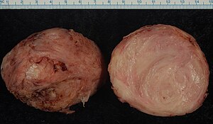
Gross examination
Examine and describe:[1]
- Location. If in the uterus:
- Intramural/submucosal/subserosal (see image)
- (Posterior/anterior/right or left lateral.)
- Number of tumors, if multiple
- Size
- (Presence or absence of any hemorrhage or necrosis.)
- ((Demarcation))
Selection
In case of hysterectomy, submit pieces from all smooth muscle tumors >5 cm in diameter.[1]
Submit any macroscopically abnormal parts of the tumors (hemorrhagic, necrotic, brittle or softening areas, and areas with blurry delimitation).[1]
Microscopic examination
Distinguish leiomyoma (benign) from leiomyosarcoma (malignant) by looking at the latter's criteria:[2]
- Marked cellular atypia
- Mitoses: > 10 mitoses/10 high power fields
- Necrosis
Diagnosis of conventional leiomyosarcoma requires 2 of these 3 histologic features.[2]
Leiomyomas typically show smooth muscle in a whorled (fascicular) pattern[3]
Leiomyosarcoma: Variable atypia, often with cytoplasmic vacuoles at both ends of nuclei, and frequent mitoses.[4]
Further information: Evaluation of tumors
Microscopic report
Report:
- Microscopic findings, including any visible linings
- Diagnosis or most probable one
- Any linings or borders.
Example, for a cervical polyp:
|
Polyp lined by a single layer of columnar epithelium consistent with endometrium. The interior consists of smooth muscles in a whorled pattern. No atypia. The finding is consistent with a pedunculated submucosal leiomyoma. |
See also: General notes on reporting
Notes
- ↑ For a full list of contributors, see article history. Creators of images are attributed at the image description pages, seen by clicking on the images. See Patholines:Authorship for details.
Main page
References
- ↑ 1.0 1.1 1.2 Monica Dahlgren, Janne Malina, Anna Måsbäck, Otto Ljungberg. Stora utskärningen. KVAST (Swedish Society of Pathology). Retrieved on 2019-09-26.
- ↑ 2.0 2.1 Paulette Mhawech-Fauceglia, M.D.. Uterus - Smooth muscle tumors - Leiomyosarcoma. Pathology Outlines. Topic Completed: 5 December 2019. Minor changes: 11 August 2020
- ↑ Mohamed Mokhtar Desouki. Uterus - Stromal tumors - Leiomyoma. Pathology Outlines. Topic Completed: 1 August 2011. Revised: 15 December 2019
- ↑ Vijay Shankar, M.D.. Soft tissue - Smooth muscle - Leiomyosarcoma - general. Pathology Outlines. Topic Completed: 1 November 2012. Revised: 11 September 2019
Image sources


