Soft tissue tumor
Author:
Mikael Häggström [note 1]
Contents
Gross processing

If it appears fatty, gross and evaluate as a lipomatous tumor.
Generally sample one slice per centimeter.
Evaluation

In case of spindle cell tumors (having elongated nuclei), the following features may help to roughly classify the tumor:
- Pointed on both ends: True fibroblastic tumors
- Pointed on one end and blunted on the other ("bullet-shaped"): Neural/nerve sheath tumors (see section below)
- Blunted on both ends ("cigar-shaped"): Smooth muscle tumor
- Triangular: Myofibroblastic
In uncertain cases, the following immunohistochemistry markers are usually helpful:
- CD34, indicating a solitary fibrous tumor
- S100, indicating a neural or nerve sheath tumor (see section below)
- Desmin, indicating a muscular tumor (skeletal muscle or Smooth muscle tumor)
- Beta catenin, indicating fibromatosis
Also consider a sarcoma as a differential diagnosis, and if unsure, have a low threshold for consulting with people with expertise in the matter, as the visual difference between benign and malignant spindle cells is relatively subtle.
Neural or nerve sheath tumors

Schwannoma, characterized by cellular Antoni A areas (top) and a loose paucicellular Antoni B areas (bottom).
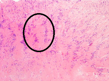
Schwannoma is also characterized by Verocay bodies (one encircled), which are stripes of anuclear zones, separated by cellular palisades.

Neurofibroma: A spindle cell lesion composed of slender fibroblast-like cells with storiform pattern and very low amount of stroma.


Further information: Evaluation of suspected malignancies
Notes
- ↑ For a full list of contributors, see article history. Creators of images are attributed at the image description pages, seen by clicking on the images. See Patholines:Authorship for details.
Main page
References
Image sources