Difference between revisions of "Stomach"
Jump to navigation
Jump to search
(Templated) |
(+Report) |
||
| Line 7: | Line 7: | ||
:*[[Gastric polyp]] | :*[[Gastric polyp]] | ||
:*[[Stomach biopsy for Helicobacter pylori]] | :*[[Stomach biopsy for Helicobacter pylori]] | ||
| − | + | {{Comprehensiveness}} | |
==Microscopic evaluation== | ==Microscopic evaluation== | ||
Generally screen for: | Generally screen for: | ||
| Line 14: | Line 14: | ||
File:Histopathology of mucosal plasma cell infiltrate in chronic gastritis.jpg|thumb|'''[[Gastritis]]''', with neutrophils indicating acute gastritis, and plasma cells (pictured) indicating chronic gastritis.<ref name=PathologyOutlinesAcute>{{cite web|url=https://www.pathologyoutlines.com/topic/stomachacutegastritis.html|title=Stomach - Gastritis - Acute gastritis|author=Elliot Weisenberg|website=pathologyOutlines}} Topic Completed: 1 August 2012. Minor changes: 31 August 2020</ref><ref>{{cite web|url=https://www.pathologyoutlines.com/topic/stomachchronicgastritis.html|title=Stomach - Gastritis - Chronic gastritis|author=Elliot Weisenberg|website=PathologyOutlines}} Topic Completed: 1 August 2012. Minor changes: 31 August 2020</ref> | File:Histopathology of mucosal plasma cell infiltrate in chronic gastritis.jpg|thumb|'''[[Gastritis]]''', with neutrophils indicating acute gastritis, and plasma cells (pictured) indicating chronic gastritis.<ref name=PathologyOutlinesAcute>{{cite web|url=https://www.pathologyoutlines.com/topic/stomachacutegastritis.html|title=Stomach - Gastritis - Acute gastritis|author=Elliot Weisenberg|website=pathologyOutlines}} Topic Completed: 1 August 2012. Minor changes: 31 August 2020</ref><ref>{{cite web|url=https://www.pathologyoutlines.com/topic/stomachchronicgastritis.html|title=Stomach - Gastritis - Chronic gastritis|author=Elliot Weisenberg|website=PathologyOutlines}} Topic Completed: 1 August 2012. Minor changes: 31 August 2020</ref> | ||
File:Histopathology of reactive gastropathy, annotated.jpg|thumb|'''Reactive gastropathy''', which is a triad of:<ref name=pmid16939055>{{Cite journal | last1 = Genta | first1 = RM. | title = Differential diagnosis of reactive gastropathy. | journal = Semin Diagn Pathol | volume = 22 | issue = 4 | pages = 273-83 | month = Nov | year = 2005 | doi = | PMID = 16939055 }}</ref><br>- '''Foveolar hyperplasia''' (black arrow), generally seen as a tortuosity in the "neck" region of the gastric glands.<br>- Scant acute and chronic '''inflammatory''' cells (white arrow).<br>- '''Smooth muscle hyperplasia''' (black oval) | File:Histopathology of reactive gastropathy, annotated.jpg|thumb|'''Reactive gastropathy''', which is a triad of:<ref name=pmid16939055>{{Cite journal | last1 = Genta | first1 = RM. | title = Differential diagnosis of reactive gastropathy. | journal = Semin Diagn Pathol | volume = 22 | issue = 4 | pages = 273-83 | month = Nov | year = 2005 | doi = | PMID = 16939055 }}</ref><br>- '''Foveolar hyperplasia''' (black arrow), generally seen as a tortuosity in the "neck" region of the gastric glands.<br>- Scant acute and chronic '''inflammatory''' cells (white arrow).<br>- '''Smooth muscle hyperplasia''' (black oval) | ||
| + | File:Helicobacter pylori, Gastric Mucosa, H&E (390307642).jpg|{{Comprehensive-begin}}Look for '''[[Helicobacter pylori]]''' even without signs of gastritis.{{Comprehensive-end}}<ref group=notes>H. pylori is very unlikely without gastritis or reactive changes.</ref> | ||
</gallery> | </gallery> | ||
| + | |||
| + | ===Microscopy report=== | ||
| + | Example in case of normal findings: | ||
| + | {|class=wikitable | ||
| + | | {{Moderate-begin}}Gastric, biopsy: {{Moderate-end}}Gastric mucosa without significant pathologic changes.<br>{{Comprehensive-begin}}Negative for helicobacter pylori organisms on H&E slide.{{Comprehensive-end}} | ||
| + | |} | ||
{{Bottom}} | {{Bottom}} | ||
Revision as of 17:18, 23 November 2020
Author:
Mikael Häggström [note 1]
| Mostly: |
Contents
Presentations
Comprehensiveness
On this resource, the following formatting is used for comprehensiveness:
- Minimal depth
- (Moderate depth)
- ((Comprehensive))
Microscopic evaluation
Generally screen for:

Gastric adenomas and adenocarcinomas.

Reactive gastropathy, which is a triad of:[3]
- Foveolar hyperplasia (black arrow), generally seen as a tortuosity in the "neck" region of the gastric glands.
- Scant acute and chronic inflammatory cells (white arrow).
- Smooth muscle hyperplasia (black oval)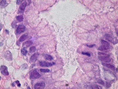
((Look for Helicobacter pylori even without signs of gastritis.))[notes 1]


.jpg)
Microscopy report
Example in case of normal findings:
| (Gastric, biopsy: )Gastric mucosa without significant pathologic changes. ((Negative for helicobacter pylori organisms on H&E slide.)) |
Notes
- ↑ H. pylori is very unlikely without gastritis or reactive changes.
- ↑ For a full list of contributors, see article history. Creators of images are attributed at the image description pages, seen by clicking on the images. See Patholines:Authorship for details.
Main page
References
- ↑ Elliot Weisenberg. Stomach - Gastritis - Acute gastritis. pathologyOutlines. Topic Completed: 1 August 2012. Minor changes: 31 August 2020
- ↑ Elliot Weisenberg. Stomach - Gastritis - Chronic gastritis. PathologyOutlines. Topic Completed: 1 August 2012. Minor changes: 31 August 2020
- ↑ Genta, RM. (Nov 2005). "Differential diagnosis of reactive gastropathy. ". Semin Diagn Pathol 22 (4): 273-83. PMID 16939055.
Image sources