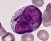
Suspected blasts on peripheral blood smear
Author:
Mikael Häggström [note 1]
When a tech or equivalent lets you know that there are suspected blasts on a peripheral blood smear, the following needs to be determined:
- If it is a new finding or a known condition in the patient, generally by a medical records lookup.
- Whether the cells are definite blast cells, suspected blast cells, or another cell type.
- The percentage of blasts compared to all white blood cells.
If it is a new finding, generally proceed as follows:
- During regular work hours, confirm that the person who reports on peripheral blood smears is notified about the case, so that the case is prioritized. Generally, the procedure is to make a complete peripheral blood smear report, in addition to calling the clinician about the presence of blasts. Further information: Peripheral blood smear
- During on call hours, ask the technician to perform a manual count if not already done.
- If you are close to the lab, generally look at the slide yourself as well to estimate the blast percentage.
- If you are far from the lab, ask a technician if images can be sent to you by micrography and telepathology.
- Consult with a senior or hematopathologist as needed, before conveying a preliminary report to the lab technician and/or clinician. At least during on call hours, it is not necessary to speculate about further sub-classification of blast cells.
Contents
Notes
- ↑ For a full list of contributors, see article history. Creators of images are attributed at the image description pages, seen by clicking on the images. See Patholines:Authorship for details.
Main page
References
Image sources
