Difference between revisions of "Template:Colorectal adenocarcinoma - microscopy criteria"
Jump to navigation
Jump to search
(→Microscopy criteria for colorectal adenocarcinoma: Specified) |
|||
| (One intermediate revision by the same user not shown) | |||
| Line 5: | Line 5: | ||
::*This should be distinguished from cases where piles of well-differentiated mucin-producing cells appear cribriform. In such piles, nuclei show regular polarity with apical mucin, and their nuclei are not markedly enlarged.<ref name=Stanford-criteria/> | ::*This should be distinguished from cases where piles of well-differentiated mucin-producing cells appear cribriform. In such piles, nuclei show regular polarity with apical mucin, and their nuclei are not markedly enlarged.<ref name=Stanford-criteria/> | ||
| − | *Invasive adenocarcinoma commonly displays: | + | *Invasive adenocarcinoma commonly displays: |
| − | :*Varying degrees of '''gland formation''' with tall columnar cells | + | :*Varying degrees of '''gland formation''' with tall columnar cells.<ref name=Stanford-criteria/> |
| − | :*Frequenty '''desmoplasia''' | + | :*Frequenty '''desmoplasia'''.<ref name=Stanford-criteria/> |
:*'''Dirty necrosis''', consisting of extensive central necrosis with granular eosinophilic karyorrhectic cell detritus.<ref name=Stanford-criteria/><ref name="LiJiang2014">{{cite journal|last1=Li|first1=Lianhuang|last2=Jiang|first2=Weizhong|last3=Yang|first3=Yinghong|last4=Chen|first4=Zhifen|last5=Feng|first5=Changyin|last6=Li|first6=Hongsheng|last7=Guan|first7=Guoxian|last8=Chen|first8=Jianxin|title=Identification of dirty necrosis in colorectal carcinoma based on multiphoton microscopy|journal=Journal of Biomedical Optics|volume=19|issue=6|year=2014|pages=066008|issn=1083-3668|doi=10.1117/1.JBO.19.6.066008}}</ref> It is located within the glandular lumina,<ref name="LiJiang2014"/> or often with a garland of cribriform glands in their vicinity.<ref name=Stanford-criteria/> | :*'''Dirty necrosis''', consisting of extensive central necrosis with granular eosinophilic karyorrhectic cell detritus.<ref name=Stanford-criteria/><ref name="LiJiang2014">{{cite journal|last1=Li|first1=Lianhuang|last2=Jiang|first2=Weizhong|last3=Yang|first3=Yinghong|last4=Chen|first4=Zhifen|last5=Feng|first5=Changyin|last6=Li|first6=Hongsheng|last7=Guan|first7=Guoxian|last8=Chen|first8=Jianxin|title=Identification of dirty necrosis in colorectal carcinoma based on multiphoton microscopy|journal=Journal of Biomedical Optics|volume=19|issue=6|year=2014|pages=066008|issn=1083-3668|doi=10.1117/1.JBO.19.6.066008}}</ref> It is located within the glandular lumina,<ref name="LiJiang2014"/> or often with a garland of cribriform glands in their vicinity.<ref name=Stanford-criteria/> | ||
| + | It may also show lymphovascular invasion. | ||
<gallery mode=packed heights=200> | <gallery mode=packed heights=200> | ||
| − | File:Micrograph of colorectal carcinoma with desmoplastic reaction.jpg| | + | File:Micrograph of colorectal carcinoma with desmoplastic reaction.jpg|'''Desmoplastic reaction''' (*) |
| − | File:Micrograph of colorectal carcinoma with dirty necrosis.jpg|Dirty necrosis | + | File:Micrograph of colorectal carcinoma with dirty necrosis.jpg|'''Dirty necrosis''' |
| + | File:Histopathology of colorectal adenocarcinoma with lymphatic invasion.jpg|thumb|'''Lymphovascular invasion''' (lymphatic invasion pictured) | ||
</gallery> | </gallery> | ||
Latest revision as of 10:38, 9 March 2021
Microscopy criteria for colorectal adenocarcinoma
- A lesion at least "high grade intramucosal neoplasia" (high grade dysplasia) has:
- Severe cytologic atypia[1]
- Cribriform architecture, consisting of juxtaposed gland lumens without stroma in between, with loss of cell polarity. Rarely, they have foci of squamous differentiation (morules).[1]
- This should be distinguished from cases where piles of well-differentiated mucin-producing cells appear cribriform. In such piles, nuclei show regular polarity with apical mucin, and their nuclei are not markedly enlarged.[1]
- Invasive adenocarcinoma commonly displays:
- Varying degrees of gland formation with tall columnar cells.[1]
- Frequenty desmoplasia.[1]
- Dirty necrosis, consisting of extensive central necrosis with granular eosinophilic karyorrhectic cell detritus.[1][2] It is located within the glandular lumina,[2] or often with a garland of cribriform glands in their vicinity.[1]
It may also show lymphovascular invasion.

Desmoplastic reaction (*)
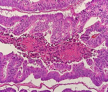
Dirty necrosis

Lymphovascular invasion (lymphatic invasion pictured)


- ↑ 1.0 1.1 1.2 1.3 1.4 1.5 1.6 Robert V Rouse. Adenocarcinoma of the Colon and Rectum. Stanford University School of Medicine. Original posting/updates: 1/31/10, 7/15/11, 11/12/11
- ↑ 2.0 2.1 Li, Lianhuang; Jiang, Weizhong; Yang, Yinghong; Chen, Zhifen; Feng, Changyin; Li, Hongsheng; Guan, Guoxian; Chen, Jianxin (2014). "Identification of dirty necrosis in colorectal carcinoma based on multiphoton microscopy ". Journal of Biomedical Optics 19 (6): 066008. doi:. ISSN 1083-3668.