Difference between revisions of "Testicle"
Jump to navigation
Jump to search
(→Evaluation: +Incidences) |
(→Evaluation: Described) |
||
| (7 intermediate revisions by the same user not shown) | |||
| Line 1: | Line 1: | ||
| − | {{Top | + | <noinclude>{{Top |
|author1=[[User:Mikael Häggström|Mikael Häggström]] | |author1=[[User:Mikael Häggström|Mikael Häggström]] | ||
|author2= | |author2= | ||
}} | }} | ||
| − | + | {{Comprehensiveness}}</noinclude> | |
==Gross processing== | ==Gross processing== | ||
===Triage=== | ===Triage=== | ||
| Line 14: | Line 14: | ||
*'''Cut''' through tunica vaginalis with scissors | *'''Cut''' through tunica vaginalis with scissors | ||
*'''Bisect''' the testis in the plane of the epididymis. | *'''Bisect''' the testis in the plane of the epididymis. | ||
| − | |||
*If any '''tumor''': | *If any '''tumor''': | ||
| + | :*'''Photograph''' the cut surface. | ||
:*Measure the '''size''' of the tumor in 3 dimensions | :*Measure the '''size''' of the tumor in 3 dimensions | ||
:*'''Describe''' the gross appearance. | :*'''Describe''' the gross appearance. | ||
| Line 23: | Line 23: | ||
*Describe the '''remaining''' testicular parenchyma. | *Describe the '''remaining''' testicular parenchyma. | ||
*If the specimen is large, serially section perpendicular to the long axis. | *If the specimen is large, serially section perpendicular to the long axis. | ||
| − | |||
===Tissue selection=== | ===Tissue selection=== | ||
| Line 39: | Line 38: | ||
Look mainly for: | Look mainly for: | ||
*Tumors. | *Tumors. | ||
| − | <gallery mode=packed> | + | <gallery mode=packed heights=230> |
| − | File:Relative incidences of testicular tumors.png|Relative incidences of testicular tumors. | + | File:Relative incidences of testicular tumors.png|Relative incidences of testicular tumors.{{MH}}<ref>{{cite journal| author=Gill MS, Shah SH, Soomro IN, Kayani N, Hasan SH| title=Morphological pattern of testicular tumors. | journal=J Pak Med Assoc | year= 2000 | volume= 50 | issue= 4 | pages= 110-3 | pmid=10851829 | doi= | pmc= | url=https://www.ncbi.nlm.nih.gov/entrez/eutils/elink.fcgi?dbfrom=pubmed&tool=sumsearch.org/cite&retmode=ref&cmd=prlinks&id=10851829 }}</ref> |
| + | File:Histopathology of seminoma.png |Classical '''seminoma''', with typical features:{{MH}}<ref>Reference for findings: {{cite web|url=https://www.pathologyoutlines.com/topic/testisseminomas.html|title=Testis & paratestis - Seminoma|author=Michelle R. Downes, M.D.|website=Pathology Outlines}} Last author update: 7 January 2020. Last staff update: 19 April 2022</ref> | ||
</gallery> | </gallery> | ||
| − | *Optionally, disorders of spermatogenesis. | + | {{Further|Tumor evaluation}} |
| + | *{{Comprehensive-begin}}Optionally, disorders of spermatogenesis.{{Comprehensive-end}} | ||
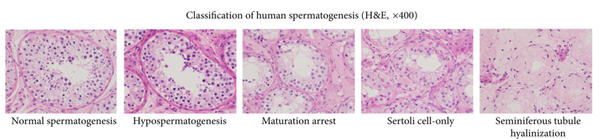
[[File:Spermatogenesis pathologies.png|850px|center]] | [[File:Spermatogenesis pathologies.png|850px|center]] | ||
| + | |||
| + | ==Reporting== | ||
| + | Example in a normal case: | ||
| + | {|class=wikitable | ||
| + | | Right testicle and spermatic cord, radical orchiectomy:<br>Benign testis{{Moderate-begin}}, negative for malignancy{{Moderate-end}}. | ||
| + | |} | ||
{{Bottom}} | {{Bottom}} | ||
Latest revision as of 16:24, 30 December 2022
Author:
Mikael Häggström [note 1]
Contents
Comprehensiveness
On this resource, the following formatting is used for comprehensiveness:
- Minimal depth
- (Moderate depth)
- ((Comprehensive))
Gross processing
Triage
Usual steps:[1]
- Weigh the specimen.
- Measure:
- The size of the testis in 3 dimensions
- The spermatic cord length and diameter
- Ink the tunica vaginalis.
- Cut through tunica vaginalis with scissors
- Bisect the testis in the plane of the epididymis.
- If any tumor:
- Photograph the cut surface.
- Measure the size of the tumor in 3 dimensions
- Describe the gross appearance.
- Measure the distance of the tumor from the tunica vaginalis
- Note tumor extension: up to, into, or through the tunica albuginea or vaginalis.
- Measure the distance of the tumor from the epididymis and spermatic cord, and note any epididymal and spermatic cord involvement.
- Describe the remaining testicular parenchyma.
- If the specimen is large, serially section perpendicular to the long axis.
Tissue selection
Generally including the following:[1]
- The proximal spermatic cord margin en face[note 2].
- For any tumor:
- At least 1 section per cm of tumor, including the closest penetration of tunica albuginea/vaginalis and epididymis.
- Grossly different areas of tumor to determine which components are present (in case of potential mixed germ cell tumor).
- Additional sections of the spermatic cord
- Normal (uninvolved) testis.
- Any found lymph nodes
In case of orchiectomy for undescended testis, submit the entire testicular parenchyma to evaluate for GCNIS (germ cell neoplasia in situ).[1]
Evaluation
Look mainly for:
- Tumors.


Further information: Tumor evaluation
- ((Optionally, disorders of spermatogenesis.))

Reporting
Example in a normal case:
| Right testicle and spermatic cord, radical orchiectomy: Benign testis(, negative for malignancy). |
Notes
- ↑ For a full list of contributors, see article history. Creators of images are attributed at the image description pages, seen by clicking on the images. See Patholines:Authorship for details.
- ↑ En face means that the section is tangential to the region of interest (such as a lesion) of a specimen. Further information: Gross_processing#Cutting
Main page
References
- ↑ 1.0 1.1 1.2 Nicole Cipriani. Testis. The University of Chicago Department of Pathology. Retrieved on 2021-03-31.
- ↑ Gill MS, Shah SH, Soomro IN, Kayani N, Hasan SH (2000). "Morphological pattern of testicular tumors. ". J Pak Med Assoc 50 (4): 110-3. PMID 10851829. Archived from the original. .
- ↑ Reference for findings: Michelle R. Downes, M.D.. Testis & paratestis - Seminoma. Pathology Outlines. Last author update: 7 January 2020. Last staff update: 19 April 2022
Image sources
- ↑ 1.0 1.1 Image(s) by: Mikael Häggström, M.D. Public Domain
- Author info
- Reusing images