Difference between revisions of "Urinary bladder"
(→Bladder cancer: Edema) |
|||
| Line 8: | Line 8: | ||
===Gross reporting of transurethral resections=== | ===Gross reporting of transurethral resections=== | ||
| − | *Generally submit '''all''' material. | + | *Generally submit '''all''' material. (It may be sufficient to submit representative sections that include the muscular layer, if grossly identified. Yet, many departments require submission of the entire specimen regardless, so if unsure, that is the safe choice.) |
| − | *Submit in tea bags or equivalent | + | *Submit in tea bags or equivalent, since tissue from transurethral resections is generally very brittle and may escape the openings of a conventional cassette. |
Example report: | Example report: | ||
Revision as of 12:15, 22 June 2022
Author:
Mikael Häggström [note 1]
Contents
Bladder cancer
The main condition of interest in urinary bladder cytologies and biopsies is bladder cancer.
Gross reporting of transurethral resections
- Generally submit all material. (It may be sufficient to submit representative sections that include the muscular layer, if grossly identified. Yet, many departments require submission of the entire specimen regardless, so if unsure, that is the safe choice.)
- Submit in tea bags or equivalent, since tissue from transurethral resections is generally very brittle and may escape the openings of a conventional cassette.
Example report:
| Container A. Labeled "bladder tumor". The specimen is received in formalin and consists of multiple fragments of tan-gray, friable soft tissue measuring about __ x __ x __ cm in aggregate. The specimen is entirely submitted for microscopic examination in __ cassettes. |
Microscopy
Mainly look for urothelial carcinoma (also called transitional cell carcinoma), which constitutes 95% of bladder cancers.[1]

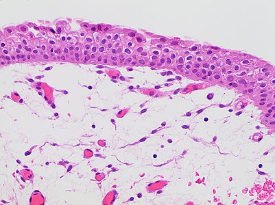
Low grade urothelial carcinoma: Urothelium is thickened but only slightly atypical and has maintained polarity.

High grade urothelial carcinoma: Loss of polarity and severe abnormal cytology.

In contrast, an inverted urothelial papilloma has smooth surface with minimal to absent exophytic component, is well circumscribed with smooth base, and has no obvious infiltration and no/minimal cytologic atypia.[2]
,_very_high_mag.jpg)
,_very_high_mag.jpg)

Other possibilities:

Squamous cell carcinoma of the urinary bladder. Further information: Urothelial versus squamous cell carcinoma
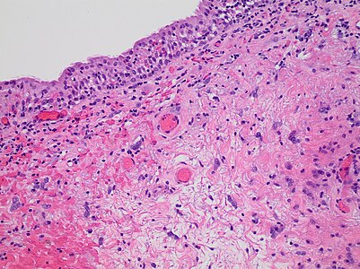
Radiation cystitis with atypical stromal cells (“radiation fibroblasts”), edema and inflammation. Check whether the patient has received radiation before making the diagnosis.
Edema (clear spaces of both the lamina propria and cytoplasm of multiple urothelial cells), which is non-specific.


Notes
- ↑ For a full list of contributors, see article history. Creators of images are attributed at the image description pages, seen by clicking on the images. See Patholines:Authorship for details.
Main page
References
- ↑ . Types of Bladder Cancer: TCC & Other Variants. CTCA.
- ↑ Monika Roychowdhury. Bladder, ureter & renal pelvis - Urothelial neoplasms - noninvasive - Inverted urothelial papilloma. Pathology Outlines. Topic Completed: 1 December 2014. Minor changes: 3 December 2020
Image sources