Emergency pathology
Author:
Mikael Häggström [note 1]
This article deals with the more time-sensitive matters encountered within pathology, and is written mainly for new pathology trainees.
Memorization-worthy:[note 2] Information relating to emergent pathology is often not conveniently and timely looked up when needed because of the need for a fast report.
Contents
Comprehensiveness
On this resource, the following formatting is used for comprehensiveness:
- Minimal depth
- (Moderate depth)
- ((Comprehensive))
Frozen sections
Even new pathology trainees may end up being the first responders to frozen sections (as well as intraoperative consultations by gross inspection only).
Preparation
Prepare at least the following:
- Finding out which senior to call for help if responding to a frozen section. A fairly new pathology trainee should generally not independently make a report to the referring physician without having at least consulted with a senior, and therefore the diagnostics of frozen section slides is not included in this section. In the meantime, ensure that you have the contact information to relevant seniors and/or attendings, and the means to perform micrography and telepathology if that person may not be able to be physically present within an acceptable time. Also consider calling additional colleagues, even at your own level or below, if there are many specimens coming at once.

- If possible, look up pertinent medical histories of potential cases that may appear as frozen sections or other forms of intraoperative consultations. For example, surgery departments may have schedules of patients for the day that you will cover frozen sections. On such lists, types of surgeries that often come as intraoperative consultations mainly include potentially malignant skin excisions, lung excisions (larger than biopsies), ovarian tumors, and samples from other common metastasis sites (lungs, liver, brain, bone). The most important details to find out are:
- What you may do yourself if you will be alone when you get the specimen, or if you need to wait for particular seniors before doing potentially irreversible steps such as inking and sectioning.
- Previous biopsies. For expected excisions from common metastasis sites, look thoroughly for any past cancer diagnoses. Note the pathologic diagnoses of the biopsies((, as well as the collection dates and accession numbers.)) (Pull out previous slides of the malignancy so that you can compare its appearance with the current case.)
- Tumor sizes where applicable(, and whether the estimation was from imaging or microscopy).
- Make sure you have a working microtome. Switch its blade if you are not certain it has had limited use.
- Prepare at least as many chucks as you think you will need according to any surgery schedule (or at least 3 of preferably various sizes).
Smear some oil on the bottom of the conductor, to avoid it getting stuck to the specimen later.[note 3]
(If you don't have a conductor, or if the embedding medium completely hardened before you applied it, perform cryotomy of the embedding medium to get a larger and smoother top.)
- Inspect the H&E staining containers and refill if necessary.
- Ensure that you have frozen section medium, as well as cover slips and mounting fluid.
Performing frozen sectioning
- Inform relevant seniors.
- Look at any requisition form or otherwise check or confirm what answers the requesting clinician wants. When the histopathologic type of tumor is known from before and its distance to the closest margin is relatively far, a gross intraoperative consultation is generally enough to measure that distance.
- Measure the size of the specimen.
- If you are permitted to proceed with the following steps, which are potentially irreversible, still consider whether there are any surprising aspects that requires you to wait until a senior arrives.
- Ink relevant margins
- 'Section the specimen so as to be able to see relevant pathologies, and closest distances to relevant margins.
- Select tissue to be able to answer the clinician's questions.
- Trim samples to make them equally thick, since you will need to section away more from thick pieces before thinner pieces will appear in cryotomy slices.
Test question
You are put on frozen sectioning duty, and a specimen arrives. You inform the attending over the phone, who will require a moment to reach the room, and therefore tells you to prepare two microscopy slides in the meantime. You make sections, but the note that instructs the number of dips or duration in the hematoxylin solution has become unreadable due to a hematoxylin stain. You don't have any phone signal in the frozen section room, so you don't have time to look it up. To what extent will you put the slide in the hematoxylin solution?
This is generally the minimal time required for proper hematoxylin staining during frozen sections. Yet, if you know the solution is not expired or diluted, and you are really in a rush, then you can possibly have it acceptably stained in 1 minute in hematoxylin. |
Troubleshooting
| Problem | Cause | Solution |
|---|---|---|
| Tissue cracks off in cryotomy |
|
Repair by warming slightly and adding embedding medium |
| Thick and thin slices |
|
|
| Wrinkled, compressed or puckered sections |
|
|
| Tissue advances but does not cut |
|
|
| Sections roll up |
|
|
| Sections thaw on the blade |
|
|
| Sections stick to the anti-roll plate or blade |
|
|
| Sections split vertically |
|
|
| Sections have fine cracks parallel to the blade edge |
|
|
| Sections show signs of vibrations |
|
|
| Stain is dull and muddy |
|
|
Thawing
Reporting of intra-operative consults
- Call the requesting clinician
- Verify that you are talking to that person, or someone who can immediately convey the diagnosis to that person. You may request to talk directly to the surgeon if a conversation is warranted.
- Tell the patient identity (with at least two different identifiers, such as name and date of birth)
- (Ask for a readback of your diagnosis. This is particularly important in cases with multiple specimens, and when it needs to be stated that a more specific diagnosis is deferred until standard slides have been evaluated.)[2]
- State your diagnosis succinctly. If necessary in cases with limited diagnostic tissue, you may state that additional tissue is required for a more certain report.
- (Make sure the clinician reads the diagnosis back to you.)
Example:
|
Frozen sectioning of suspected malignant skin excisions
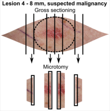
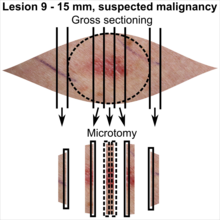
While most frozen sections can be predicted from schedules of the operating room and thereby be looked up beforehand, suspected malignant skin excisions often come from outside the main surgery department, even more indicating memorization of how to handle them.
Tissue selection
| <4 mm | 4 - 8 mm | 9 - 15 mm |
|---|---|---|

|

|

|
In table above, each top image shows recommended lines for cutting out slices to be submitted for further processing. Bottom image shows which side of the slice that should be put to microtomy. Dashed lines here mean that either side could be used. Further information: Gross processing of skin excisions
Donor kidney biopsy
Gross processing
Prepare a separate slide for right versus left kidney if applicable.
Microscopic evaluation
Generally evaluate as per local protocol, which generally includes the following parameters, given individually for right and left kidney:
Other frozen sections
Although these are generally given on schedules of the operating room, any pathologist may end up suddenly covering for another one, and subsequently be presented with the frozen section case without having had the time to look it up beforehand.
In malignant or suspected malignant cases, surgical margins are generally more important than main tumor diagnosis.
- In lung wedge resection and lobectomy, sample the surgical margin that is presumably closest to a tumor for frozen sectioning. Further information: Lung wedge resection and lobectomy
- In an intestine with tumor, it is generally enough to open the intestine (avoiding cutting through a tumor unless it is corcumferantial, and grossly measuring the distances to the proximal and distal surgical margins, as well as the distances to the radial or mesenteric surgical margins. Further information: Intestine with tumor
- For a donor kidney biopsy, generally evaluate as per local protocol. Further information: Donor kidney biopsy
However, for myocarditis requests (see the heart article), generally have the cardiac biopsy sent in formalin for regular H&E processing (even on weekends) rather than perform frozen sectioning, since good quality slides is the priority.
See also
Notes
- ↑ For a full list of contributors, see article history. Creators of images are attributed at the image description pages, seen by clicking on the images. See Patholines:Authorship for details.
- ↑ Further information on what is memorization-worthy or not: Learning pathology
- ↑ Do not smear oil on the bottom of the conductor before placing it on a chuck without a specimen, for chuck preparation, as it may (theoretically) cause breakage of the specimen later because of slippage of a segment over the oil layer.
- ↑ The excision example shows a superficial basal cell carcinoma.
Main page
References
- ↑ A list of included entries and references is found on main image page in Wikimedia Commons: Commons:File:Metastasis sites for common cancers.svg#Summary
- ↑ Wiggett A, Fischer G (2023). "Intraoperative Communications Between Pathologists and Surgeons: Do We Understand Each Other? ". Arch Pathol Lab Med 147 (8): 933-939. doi:. PMID 36343374. Archived from the original. .
- ↑ There are many variants for the processing of skin excisions. These examples use aspects from the following sources:
- . Handläggning av hudprover – provtagningsanvisningar, utskärningsprinciper och snittning (Handling of skin samples - sampling instructions, cutting principles and incision. Swedish Society of Pathology.
- For number of slices and coverage of lesions, depending on size. - Monica Dahlgren, Janne Malina, Anna Måsbäck, Otto Ljungberg. Stora utskärningen. KVAST (Swedish Society of Pathology). Retrieved on 2019-09-26.
- For slices towards the pointy ends to determine radicality, which can be parallel to the slices through the lesions (shown), or as longitudinal slices that go through each pointy end. - . Dermatopathology Grossing Guidelines. University of California, Los Angeles. Retrieved on 2019-10-23.
- For microtomy of the most central side at the lesion - "The principles of mohs micrographic surgery for cutaneous neoplasia
- With a "standard histologic examination" that, in addition to the lesion, only includes one section from each side along the longest diameter of the specimen.
- It also shows an example of circular coverage, with equal coverage distance in all four directions.
- The entire specimen may be submitted if the risk of malignancy is high. - . Handläggning av hudprover – provtagningsanvisningar, utskärningsprinciper och snittning (Handling of skin samples - sampling instructions, cutting principles and incision. Swedish Society of Pathology.
- ↑ Schelling JR (2016). "Tubular atrophy in the pathogenesis of chronic kidney disease progression. ". Pediatr Nephrol 31 (5): 693-706. doi:. PMID 26208584. PMC: 4726480. Archived from the original. .
Image sources
- ↑ Image(s) by: Mikael Häggström, M.D. Public Domain
- Author info
- Reusing images


