Pancreatic tumor
Jump to navigation
Jump to search
Author:
Mikael Häggström [note 1]
Microscopic evaluation
If a biopsy has been processed both for histology and cytology, make sure that their results are not contradictory.
Stages of pancreatic intraepithelial neoplasia.[1]
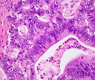
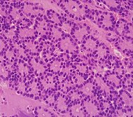
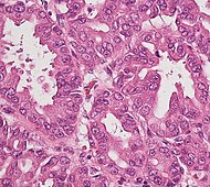
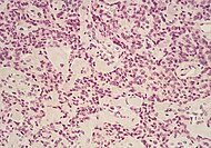
Relative incidences of various pancreatic neoplasm.[2]
| Cancer type | Relative incidence[3] | Microscopy findings[3] | Micrograph | ||
|---|---|---|---|---|---|
| Pancreatic ductal adenocarcinoma (PDAC) | 90% | Glands and desmoplasia | 
| ||
| Pancreatic acinar cell carcinoma (ACC) | 1% to 2% | Granular appearance | 
| ||
| Solid pseudopapillary tumor | Discohesive tumor nests surrounded by thin fibrous bands. |  Low and high magnification[4] | |||
| Adenosquamous carcinoma | 1% to 4%[5] | epithelial cells. | 
| ||
| Pancreatic neuroendocrine tumor | 5% | Multiple nests of tumor cells | Gastrinoma | ||
| Pre-cancer below for comparison: | |||||
| Precancer: Intraductal papillary mucinous neoplasm (IPMN) |
3% | Mucinous epithelial cells.[6] Growth within the pancreatic ducts.[7] | 
| ||
Staging
Stage pancreatic cancers as follows:[8]
| Tumor (T) | Criteria |
|---|---|
| TX | The primary tumor cannot be evaluated. |
| T0 | No evidence of cancer was found in the pancreas. |
| Tis | Carcinoma in situ. This includes:
|
| T1 | The tumor is in the pancreas only, and is ≤2 cm in greatest dimension |
| - T1a | Tumor ≤0.5 cm in greatest dimension |
| - T1b | Tumor >0.5 cm and <1 cm in greatest dimension |
| - T1c | Tumor 1–2 cm in greatest dimension |
| T2 | The tumor is in the pancreas only, and it is >2 cm and ≤4 cm in greatest dimension |
| T3 | Tumor >4 cm in greatest dimension |
| T4 | Tumor involves celiac axis, superior mesenteric artery, and/or common hepatic artery, regardless of size |
| Node (N) | Criteria |
|---|---|
| NX | The regional lymph nodes cannot be evaluated. |
| N0 | Cancer was not found in the regional lymph nodes. |
| N1 | Metastasis in 1 to 3 regional lymph nodes. |
| N2 | Metastasis in 4 or more regional lymph nodes. |
| Metastasis (M) | Criteria |
|---|---|
| M0 | No distant metastasis |
| M1 | Distant metastasis, including distant lymph nodes. Pancreatic cancer most commonly spreads to the liver, the peritoneum, and the lungs. |
Further information: Evaluation of tumors
Notes
- ↑ For a full list of contributors, see article history. Creators of images are attributed at the image description pages, seen by clicking on the images. See Patholines:Authorship for details.
Main page
References
- ↑ Hackeng WM, Hruban RH, Offerhaus GJ, Brosens LA (2016). "Surgical and molecular pathology of pancreatic neoplasms.
". Diagn Pathol 11 (1): 47. doi:. PMID 27267993. PMC: 4897815. Archived from the original. .
- "This article is distributed under the terms of the Creative Commons Attribution 4.0 International License (http://creativecommons.org/licenses/by/4.0/)"
- Image title and optimization: Mikael Häggström, M.D. - ↑ Diagram by Mikael Häggström, M.D.
Source data: Wang Y, Miller FH, Chen ZE, Merrick L, Mortele KJ, Hoff FL (2011). "Diffusion-weighted MR imaging of solid and cystic lesions of the pancreas. ". Radiographics 31 (3): E47-64. doi:. PMID 21721197. Archived from the original. . - ↑ 3.0 3.1 Unless otherwise specified in boxes, reference is: "Therapeutic Implications of Molecular Subtyping for Pancreatic Cancer ". Oncology 31 (3): 159–66, 168. March 2017. PMID 28299752. Archived from the original. .
- ↑ Image by Mikael Häggström, MD.
Reference for features: Pooja Navale, M.D., Omid Savari, M.D., Joseph F. Tomashefski, Jr., M.D., Monika Vyas, M.D.. Solid pseudopapillary neoplasm. Last author update: 4 March 2022 - ↑ "Adenosquamous carcinoma of the pancreas: a case report ". Cases Journal 3 (1): 41. February 2010. doi:. PMID 20205828.
- ↑ Diana Agostini-Vulaj. Pancreas – Exocrine tumors / carcinomas – Intraductal papillary mucinous neoplasm (IPMN). Pathology Outlines. Topic Completed: 1 July 2018. Revised: 9 March 2020
- ↑ "Pathologic Evaluation and Reporting of Intraductal Papillary Mucinous Neoplasms of the Pancreas and Other Tumoral Intraepithelial Neoplasms of Pancreatobiliary Tract: Recommendations of Verona Consensus Meeting ". Annals of Surgery 263 (1): 162–77. January 2016. doi:. PMID 25775066.
- ↑ Amin, Mahul (2017). AJCC cancer staging manual
(8 ed.). Switzerland: Springer. ISBN 978-3-319-40617-6. OCLC 961218414.
- For access, see the Secrets chapter of Patholines.
- Copyright note: The AJCC, 8th Ed. is published by a company in Switzerland, and the tables presented therein are Public Domain because they consist of tabular information without literary or artistic innovation, and therefore do not fulfil the inclusion criterion of the Swiss Copyright Act (CopA) which applies to "literary and artistic intellectual creations with individual character" (see Federal Act on Copyright and Related Rights (Copyright Act, CopA) of 9 October 1992 (Status as of 1 January 2022)).
Image sources


