Prostate adenocarcinoma
Author:
Mikael Häggström [note 1]
Comprehensiveness
On this resource, the following formatting is used for comprehensiveness:
- Minimal depth
- (Moderate depth)
- ((Comprehensive))
Gross processing
As prostatectomy or biopsy.
Microscopic evaluation
Screening method
- Before microscopy, look at each microscopy slide by eye, to plan the microscopy screening so as to not miss peripheral fragments.
- Screen at low power, and switch to high power when encountering glandular structures that can not otherwise be cleared. Look in particular for those surrounding nerves.
- At least if no cancer is seen, also look for inflammation.[notes 1]
Characteristics of adenocarcinoma
- Relatively common and highly specific findings of prostatic adenocarcinoma
- [1]
-
 Multiple nucleoli (Pictured in an acinar adenocarcinoma, the most common subdiagnosis of prostate adenocarcinoma)
 -
![Eccentric nucleoli[1] (pictured example has double and eccentric nucleoli).](https://upload.wikimedia.org/wikipedia/commons/thumb/8/8f/Micrograph_of_acinar_adenocarcinoma_of_the_prostate_with_double_and_marginated_nucleoli.jpg/330px-Micrograph_of_acinar_adenocarcinoma_of_the_prostate_with_double_and_marginated_nucleoli.jpg) Eccentric nucleoli[1] (pictured example has double and eccentric nucleoli).
Eccentric nucleoli[1] (pictured example has double and eccentric nucleoli).

![Eccentric nucleoli[1] (pictured example has double and eccentric nucleoli).](/File:Micrograph_of_acinar_adenocarcinoma_of_the_prostate_with_double_and_marginated_nucleoli.jpg)
- Specific but relatively rare signs of adenocarcinoma
- [notes 2]
![Perineural invasion.[1] It should be circumferential (as pictured) to count.[2]](/File:Histopathology_of_prostatic_adenocarcinoma_with_circumferential_perineural_invasion.jpg)
On biopsies, look in particular near the tips for perineural invasion, as it is most likely seen by the capsule. Glands adjacent to and indenting nerves is not sufficient as a diagnostic criterion by itself. Glands partially surrounding a nerve is an indication of carcinoma.[2]
- Collagenous micronodules for acinar adenocarcinoma[1]
- Angiolymphatic invasion[1]
- Extraprostatic extension,[1] which in biopsies can be diagnosed when tumor cells are located in fatty tissue.
- Less specific findings
-
![Mitoses: also seen in for example high-grade prostatic intraepithelial neoplasia (HGPIN) and prostate inflammation.[1] Picture shows adenocarcinoma with two mitoses in reactive epithelium.](https://upload.wikimedia.org/wikipedia/commons/thumb/6/64/Micrograph_of_adenocarcinoma_of_the_prostate_with_two_mitoses_in_reactive_epithelium.jpg/330px-Micrograph_of_adenocarcinoma_of_the_prostate_with_two_mitoses_in_reactive_epithelium.jpg) Mitoses: also seen in for example high-grade prostatic intraepithelial neoplasia (HGPIN) and prostate inflammation.[1] Picture shows adenocarcinoma with two mitoses in reactive epithelium.
Mitoses: also seen in for example high-grade prostatic intraepithelial neoplasia (HGPIN) and prostate inflammation.[1] Picture shows adenocarcinoma with two mitoses in reactive epithelium. -
![Intraluminal blue mucin[1] (pictured in acinar adenocarcinoma)](https://upload.wikimedia.org/wikipedia/commons/thumb/8/86/Micrograph_of_acinar_adenocarcinoma_of_the_prostate_with_blue_mucin.jpg/330px-Micrograph_of_acinar_adenocarcinoma_of_the_prostate_with_blue_mucin.jpg) Intraluminal blue mucin[1] (pictured in acinar adenocarcinoma)
Intraluminal blue mucin[1] (pictured in acinar adenocarcinoma) -
![Intraluminal atypical eosinophilic secretions.[1]](https://upload.wikimedia.org/wikipedia/commons/thumb/0/03/Histopathology_of_prostatic_adenocarcinoma_with_atypical_eosinophilic_secretions.jpg/330px-Histopathology_of_prostatic_adenocarcinoma_with_atypical_eosinophilic_secretions.jpg) Intraluminal atypical eosinophilic secretions.[1]
Intraluminal atypical eosinophilic secretions.[1] -
![Intraluminal crystalloids.[3]](https://upload.wikimedia.org/wikipedia/commons/thumb/5/59/Histopathology_of_prostatic_intraluminal_crystalloid.jpg/500px-Histopathology_of_prostatic_intraluminal_crystalloid.jpg) Intraluminal crystalloids.[3]
Intraluminal crystalloids.[3] -
 Uneven distribution and infiltrative pattern of glands
 -
![Glomerulations, for acinar adenocarcinoma, consisting of epithelial proliferations into one or more gland lumina, typically a cribriform tuft with a single attachment to the gland wall.[2]](https://upload.wikimedia.org/wikipedia/commons/c/c3/Micrograph_of_prostate_adenocarcinoma_with_a_glomeruloid_gland.jpg) Glomerulations, for acinar adenocarcinoma, consisting of epithelial proliferations into one or more gland lumina, typically a cribriform tuft with a single attachment to the gland wall.[2]
Glomerulations, for acinar adenocarcinoma, consisting of epithelial proliferations into one or more gland lumina, typically a cribriform tuft with a single attachment to the gland wall.[2]
![Mitoses: also seen in for example high-grade prostatic intraepithelial neoplasia (HGPIN) and prostate inflammation.[1] Picture shows adenocarcinoma with two mitoses in reactive epithelium.](/File:Micrograph_of_adenocarcinoma_of_the_prostate_with_two_mitoses_in_reactive_epithelium.jpg)
![Intraluminal blue mucin[1] (pictured in acinar adenocarcinoma)](/File:Micrograph_of_acinar_adenocarcinoma_of_the_prostate_with_blue_mucin.jpg)
![Intraluminal atypical eosinophilic secretions.[1]](/File:Histopathology_of_prostatic_adenocarcinoma_with_atypical_eosinophilic_secretions.jpg)
![Intraluminal crystalloids.[3]](/File:Histopathology_of_prostatic_intraluminal_crystalloid.jpg)

![Glomerulations, for acinar adenocarcinoma, consisting of epithelial proliferations into one or more gland lumina, typically a cribriform tuft with a single attachment to the gland wall.[2]](/File:Micrograph_of_prostate_adenocarcinoma_with_a_glomeruloid_gland.jpg)
- Prominent nucleoli[1]
- Nuclear enlargement
Precancerous lesions
,_annotated.jpg)
In case of only less specific findings, consider a Prostatic intraepithelial neoplasia (PIN) or an atypical small acinar proliferation (ASAP).
A PIN is where acini are architecturally benign, but individual cells display atypia. In high-grade PIN (HGPIN), the changes are similar to those of prostate cancer, whereas in low-grade (LGPIN) the changes are milder. Most pathologists do not report the presence of LGPIN.[5]
An ASAP is a lesion that is probably carcinoma but either lacks definitive diagnostic features, or is too small to be certain (see image below).[6] It should not be used for benign lesions that are just unusual looking.[6] In uncertain cases, a diagnosis of adenocarcinoma can be excluded by immunohistochemical detection of basal cells (or confirmed by absence thereof),[1] such as using the PIN-4 cocktail of stains (which consists of P504S, p63 and high-molecular-weight keratins (HMWK) such as CK5 and CK14).
-
 Small acinar cell proliferation, with acinar cells with large nuclei, prominent nucleoli (arrows marking two of them) and no certain basal cell lining.
 -
 PIN-4 staining of benign prostate gland and adenocarcinoma
.jpg)

Picture above compares a PIN-4 immunohistochemistry of benign gland (left) and adenocarcinoma (right) using PIN-4. The adenocarcinoma lacks the basal epithelial cells (stained dark brown by p63 and HMWK). Also, in PIN-4 stained samples, adenocarcinoma cells generally display red cytoplasms (stained by AMACR, also known as P504S), while benign glands do not.
-
 Atrophy is a differential diagnosis to prostate cancer. This example shows gradually increasing simple atrophy from left to right, H&E stain. Crowding and angulation may mimic that of adenocarcinoma, but there is nuclear basophilia rather than atypia, and occasional basal cells can still be seen.
 -
![Also, seminal vesicle glands may mimic prostatic adenocarcinoma by crowded glands with enlarged hyperchromatic and irregular nuclei, but will have inconspicuous nucleoli and coarse refractile golden brown lipofuscin granules.[7]](https://upload.wikimedia.org/wikipedia/commons/thumb/d/d2/Histology_of_seminal_vesicle_glands_on_H%26E_stain.jpg/500px-Histology_of_seminal_vesicle_glands_on_H%26E_stain.jpg) Also, seminal vesicle glands may mimic prostatic adenocarcinoma by crowded glands with enlarged hyperchromatic and irregular nuclei, but will have inconspicuous nucleoli and coarse refractile golden brown lipofuscin granules.[7]
Also, seminal vesicle glands may mimic prostatic adenocarcinoma by crowded glands with enlarged hyperchromatic and irregular nuclei, but will have inconspicuous nucleoli and coarse refractile golden brown lipofuscin granules.[7]

![Also, seminal vesicle glands may mimic prostatic adenocarcinoma by crowded glands with enlarged hyperchromatic and irregular nuclei, but will have inconspicuous nucleoli and coarse refractile golden brown lipofuscin granules.[7]](/File:Histology_of_seminal_vesicle_glands_on_H%26E_stain.jpg)
Subdiagnoses

The histopathologic subdiagnosis of prostate cancer has implications for the possibility and methodology of any subsequent Gleason scoring.[8] The most common histopathological subdiagnosis of prostate cancer is acinar adenocarcinoma, constituting 93% of prostate cancers.[9] The most common form of acinar adenocarcinoma, in turn, is "adenocarcinoma, not otherwise specified", also termed conventional, or usual acinar.[10]
| Subdiagnosis | Relative incidence | Image | Microscopic characteristics | Immunohistochemistry | Gleason scoring | ||
|---|---|---|---|---|---|---|---|
| Core biopsy |
Radical prostatectomy | ||||||
| Acinar adenocarcinoma - 93%[9] |
Adenocarcinoma (not otherwise specified/ conventional/ usual acinar)[10] |
77%[notes 4] | 54%[notes 4] | _with_glomeruloid_glands.jpg)
|
Tumorous glands: | As usual | |
| Foamy gland carcinoma | 17%[11][notes 3] | 13–23%[11][notes 3] | Based on architecture, discounting foamy cytoplasms[8] | ||||
| Atrophic carcinoma | 2%[11][notes 5] | 16%[11][notes 5] | Tumorous glands: | As usual[8] | |||
| Pseudohyperplastic carcinoma | 2%[11] | 11%[11] |
|
Tumorous glands: | 3+3=6[8] | ||
| Microcystic carcinoma | 11%[11] | On (usually) adjacent acinar adeocarcinoma[12] | |||||
| PIN-like | 1.3%[13] |
|
Tumorous glands:
|
Not recommended[8] | |||
| Non acinar (or mixed acinar/ non-acinar) adenocarcinoma |
Ductal adenocarcinoma | 3% to 12.7%[14][notes 3] | 
|
||||
| Intraductal adenocarcinoma | 2.8%[16] |  H&E and CK5/6 |
|||||
| Urothelial carcinoma | 0.7 to 2.8%[18] | 
|
Not recommended[8] | ||||
| Small-cell carcinoma | 0.3–2%[20][21][notes 3] | Error creating thumbnail: |
Half of cases have usual acinar components[8] |
||||
| Mucinous adenocarcinoma | 0.2%[18] | File:Micrograph of mucinous adenocarcinoma of the prostate with Gleason score 7 (3 + 4) with individual well-formed glands and minor component of cribriform glands floating in extracellular mucin.jpg |
|
Tumorous glands: | 4+4=8 for irregular cribriform glands floating in mucin.[8] | ||
| Signet-ring adenocarcinoma | 0.02%[22] | File:Micrograph of signet-ring adenocarcinoma of the prostate.jpg |
|
Tumorous glands: | Not recommended[8] | ||
| Basal-cell carcinoma | 0.01%[23] | Basaloid tumor:
BCC-pattern: |
Not recommended.[8] | ||||
Gleason scoring
Rate the dominant, or most common cell morphology (scored 1—5), in addition to the non-dominant cell pattern with the highest grade (scored 1—5).
-
 Gleason pattern 6 (3+3).jpg
 -
 Gleason pattern 7 (3+4).jpg
 -
 Gleason pattern 8 (4+4).jpg
 -
 Gleason pattern 9 (4+5).jpg
 -
 Gleason pattern 10 (5+5).jpg
.jpg)
.jpg)
.jpg)
.jpg)
.jpg)
Gleason score is as follows:[24]
- Gleason score ≤3: Only individual discrete well-formed glands
- Gleason score 4: Poorly-formed, fused and/or cribriform glands
- Gleason score 5: Lacks gland formation, or has necrosis
-
 Gleason score 6 (3+3)
 -
 Cribriform pattern: Gleason grade 4
 -
 Gleason score 7 (3+4) with minor component of cribriform glands
 -
 Gleason score 8 (4+4) with glomeruloid glands
 -
 Gleason score 8 (4+4) with irregular cribriform glands
 -
 Gleason score 8 (4+4) with fused glands with cytoplasmic vacuoles
 -
 Gleason score 8 (4+4) with poorly-formed glands
 -
 Gleason score 9 (4+5) with cribriform glands, some with necrosis
 -
 Gleason score 10 (5+5) with cords of cells
 -
 Gleason score 10 (5+5) with individual cells
 -
 Gleason score 10 (5+5) with solid sheets of cells
.jpg)

_with_minor_component_of_cribriform_glands.jpg)
_with_irregular_cribriform_glands.jpg)
_with_fused_glands_with_cytoplasmic_vacuoles.jpg)
_with_poorly-formed_glands.jpg)
_with_cribriform_glands,_some_with_necrosis.jpg)
_with_cords_of_cells.jpg)
_with_individual_cells.jpg)
_with_solid_sheets_of_cells.jpg)
_with_individual_well-formed_glands_and_minor_component_of_cribriform_glands_floating_in_extracellular_mucin.jpg)
The percentage of pattern 4 should be reported for Gleason score 3+4 ((and 4+3)).[25]
In case of 3 patterns in one specimen:[26]
- On a biopsy: most common pattern + the highest pattern. For example, mostly pattern 3, a bit of 4, and a tiny bit of 5 would be graded 3 + 5 = 8.
- On a prostatectomy: First and second most prevalent patterns, and if there is a minor or third component of higher grade, that is termed a tertiary pattern. The same example as above would be classified as 3 + 4 = 7 with tertiary pattern 5.
((In cases of Gleason pattern 3+4 and 4+3, also state the percentage of Gleason pattern 4 compared to the entire tumor area.[27]))
Grade group
(In a moderately comprehensive report, also state the overall grade group for the adenocarcinoma:[24]
- Grade group 1 (Gleason score ≤6) - Only individual discrete well-formed glands
- Grade group 2 (Gleason score 3+4=7) - Predominantly well-formed glands with a lesser component of poorly-formed, fused and/or cribriform glands
- Grade group 3 (Gleason score 4+3=7) - Predominantly poorly-formed, fused, and/or cribriform glands with a lesser component of well-formed glands
- Grade group 4 (Gleason score 8), either of the following
- Only poorly-formed, fused and/or cribriform glands
- Predominantly well-formed glands with a lesser component that lacks glands
- Predominantly lacking glands with a lesser component of well-formed glands
- Grade group 5 (Gleason scores 9-10): Any area that lacks gland formation, or with necrosis)
Staging
Depending on sample type:
- Multiple biopsy specimens: Adenocarcinoma presence in how many of the biopsies
- Prostatectomy: Stage by TNM:
The AJCC 8th edition[28] has a shorter pathological tumor classification (pT) than the clinical one (cT). pT does not have any T1 category, and is as follows:
- Evaluation of the (primary) tumor ('T')
- T2: Organ confined
- T3: Extraprostatic extension
- T3a: Extraprostatic extension (unilateral or bilateral) or microscopic invasion of bladder neck
- T3b: Tumor invades seminal vesicle(s)
- T4: Tumor is fixed or invades adjacent structures other than seminal vesicles such as external sphincter, rectum, bladder, levator muscles, and/or pelvic wall
- Evaluation of the regional lymph nodes ('N')
- NX: Regional lymph nodes cannot be assessed
- N0: No positive regional nodes
- N1: Metastases in regional node(s)
- Evaluation of distant metastasis ('M')
- M0: No distant metastasis
- M1: Distant metastasis
- M1a: Nonregional lymph node(s)
- M1b: Bone(s)
- M1c: Other site(s) with or without bone disease
When more than one site of metastasis is present, the most advanced category is used. For cancers, generally include a synoptic report, such as per College of American Pathologists (CAP) protocols at cap.org/protocols-and-guidelines.
- Further information: Evaluation
Reporting
- Diagnosis
- Gleason score, and optionally grade group
- Extent
- Prostatectomy: Stage.
- Prostate biopsies: Number of biopsies where tumor is found
- Both prostatectomy and biopsies: Percentage of tumor involvement (see Percentage section below).
- Any perineural or angiolymphatic invasion.
- ((Any inflammation.[notes 1]))
Examples in a biopsy with adenocarcinoma:
| (Prostate, left apex, needle biopsy:) Prostatic adenocarcinoma, Gleason score 3+3 = 6 (grade group 1), involving 30% of one out of one core. |
If the microscopy and gross description give discordant number of cores, you may use the gross number.
Prostate, right apex, needle biopsy:
|
Template for prostatectomy:
Prostate and seminal vesicles, radical robotic prostatectomy:
|
For cancers, generally include a synoptic report, such as per College of American Pathologists (CAP) protocols at cap.org/protocols-and-guidelines.
Percentage
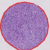
In prostatectomies, percentage of involvement can be estimated by marking the tumor edges with a marker pen under the microscope, and looking at the slide without microscope to roughly estimate the percentage of involvement. The total percentage can be calculated as the average of the involvement of each slide.
In biopsies, percentage of involvement can be estimated by first estimating what percentage of the field-of-view diameter the tumorous area occupies, or how many field-of-view diameters it occupies, divided by how many field-of-view diameters the entire biopsy occupies. When there is at least 1 mm of non-involved segment between separate tumor foci in a biopsy, give the percentage as if the tumor involved such segments too, but denote it as "discontinuously" involving it.
See also: General notes on reporting
Notes
- ↑ 1.0 1.1 Inflammation can explain for example a high PSA value in the absence of adenocarcinoma, so its reporting is usually only needed in such cases.
- ↑ "Rare" here refers to prevalence at least in core biopsies.(Cruz 2016)
- ↑ 3.0 3.1 3.2 3.3 3.4 At least where noted, the numbers include cases where the pattern is found admixed with usual acinar adenocarcinoma.
- ↑ 4.0 4.1 Numbers for usual acinar adenocarcinoma do not include mixed patterns with other subdiagnoses.
- ↑ 5.0 5.1 Number refers to sporadic atrophic pattern adenocarcinoma.
- ↑ For a full list of contributors, see article history. Creators of images are attributed at the image description pages, seen by clicking on the images. See Patholines:Authorship for details.
Main page
References
- ↑ 1.00 1.01 1.02 1.03 1.04 1.05 1.06 1.07 1.08 1.09 1.10 1.11 1.12 Cruz, Andrea O.; Santana, Amanda L. S.; Santos, Andréia C.; Athanazio, Daniel A. (2016). "Frequency of the morphological criteria of prostate adenocarcinoma in 387 consecutive prostate needle biopsies: emphasis on the location and number of nucleoli
". Jornal Brasileiro de Patologia e Medicina Laboratorial. doi:. ISSN 1676-2444.
Attribution 4.0 International (CC BY 4.0) license - ↑ 2.0 2.1 2.2 Robert V Rouse MD. Prostatic Adenocarcinoma. Stanford Medical School. Last update 2/2/16
- ↑ Svatek, R S; Karam, J A; Rogers, T E; Shulman, M J; Margulis, V; Benaim, E A (2007). "Intraluminal crystalloids are highly associated with prostatic adenocarcinoma on concurrent biopsy specimens ". Prostate Cancer and Prostatic Diseases 10 (3): 279–282. doi:. ISSN 1365-7852.
- ↑ Image by Mikael Häggström, MD. Reference for features:
- Margaret Sanders, M.B.B.Ch., Murali Varma, M.B.B.S.. High grade prostatic intraepithelial neoplasia (HGPIN). Pathology Outlines. Last author update: 23 February 2021 - ↑ Stanley A Brosman, MD. Precancerous Lesions of the Prostate. Medscape. Updated: Feb 26, 2020
- ↑ 6.0 6.1 . Prostatic Adenocarcinoma - Atypical Small Acinar Proliferation (ASAP). Stanford Medical School. Retrieved on 2020-09-14.
- ↑ Image by Mikael Häggström, MD. Reference for findings: Faryal Shoaib, M.D., Chinedum Okafor, M.D., Y. Albert Yeh, M.D., Ph.D.. Anatomy & histology-seminal vesicles / ejaculatory duct. Pathology Outlines. Last staff update: 20 November 2023
- ↑ 8.00 8.01 8.02 8.03 8.04 8.05 8.06 8.07 8.08 8.09 8.10 8.11 8.12 8.13 8.14 8.15 8.16 8.17 8.18 8.19 8.20 8.21 8.22 8.23 8.24 8.25 8.26 8.27 8.28 8.29 8.30 8.31 8.32 8.33 8.34 8.35 8.36 8.37 8.38 8.39 8.40 8.41 8.42 8.43 8.44 8.45 8.46 8.47 8.48 8.49 8.50 8.51 8.52 8.53 8.54 8.55 8.56 8.57 8.58 8.59 8.60 "The pathology of unusual subtypes of prostate cancer ". Chin. J. Cancer Res. 28 (1): 130–43. February 2016. doi:. PMID 27041935.
- ↑ 9.0 9.1 9.2 9.3 9.4 9.5 Baig, Faraz A.; Hamid, Amna; Mirza, Talat; Syed, Serajuddaula (2015). "Ductal and Acinar Adenocarcinoma of Prostate: Morphological and Immunohistochemical Characterization ". Oman Medical Journal 30 (3): 162–166. doi:. ISSN 1999768X.
- ↑ 10.0 10.1 . Prostatic Adenocarcinoma. Stanford University School of Medicine. Retrieved on 2019-10-30.
- ↑ 11.0 11.1 11.2 11.3 11.4 11.5 11.6 Humphrey, Peter A (2018). "Variants of acinar adenocarcinoma of the prostate mimicking benign conditions ". Modern Pathology 31 (S1): 64–70. doi:. ISSN 0893-3952.
- ↑ 12.0 12.1 12.2 12.3 12.4 12.5 Yaskiv, Oksana; Cao, Dengfeng; Humphrey, Peter A. (2010). "Microcystic Adenocarcinoma of the Prostate: A Variant of Pseudohyperplastic and Atrophic Patterns ". The American Journal of Surgical Pathology 34 (4): 556–561. doi:. ISSN 0147-5185.
- ↑ Zhou, Ming (2018). "High-grade prostatic intraepithelial neoplasia, PIN-like carcinoma, ductal carcinoma, and intraductal carcinoma of the prostate ". Modern Pathology 31 (S1): 71–79. doi:. ISSN 0893-3952.
- ↑ "The update of prostatic ductal adenocarcinoma ". Chin. J. Cancer Res. 28 (1): 50–7. February 2016. doi:. PMID 27041926.
- ↑ Robert V Rouse (2012-01-06). Prostatic Ductal Adenocarcinoma. Stanford University School of Medicine.
- ↑ 16.0 16.1 16.2 16.3 Magers, Martin; Kunju, Lakshmi Priya; Wu, Angela (2015). "Intraductal Carcinoma of the Prostate: Morphologic Features, Differential Diagnoses, Significance, and Reporting Practices ". Archives of Pathology & Laboratory Medicine 139 (10): 1234–1241. doi:. ISSN 0003-9985.
- ↑ 17.0 17.1 Roberts, Jordan A.; Zhou, Ming; Park, Yong Wok; Ro, Jae Y. (2013). "Intraductal Carcinoma of Prostate: A Comprehensive and Concise Review ". Korean Journal of Pathology 47 (4): 307. doi:. ISSN 1738-1843.
- ↑ 18.0 18.1 Grignon, David J (2004). "Unusual subtypes of prostate cancer ". Modern Pathology 17 (3): 316–327. doi:. ISSN 0893-3952.
- ↑ 19.0 19.1 19.2 Robert V Rouse. Papillary Urothelial (Transitional Cell) Carcinoma. Stanford University School of Medicine. Original posting/updates: 10/20/12, 12/29/12
- ↑ 0.3%-1%: Page 77 in:Beltran, Antonio (2017). Pathology of the prostate : an algorithmic approach . Cambridge, United Kingdom New York, NY: Cambridge University Press. ISBN 978-1-108-18565-3. OCLC 1011514854.
- ↑ 0.5-2%: Kumar, Kishore; Ahmed, Rafeeq; Chukwunonso, Chime; Tariq, Hassan; Niazi, Masooma; Makker, Jasbir; Ihimoyan, Ariyo (2018). "Poorly Differentiated Small-Cell-Type Neuroendocrine Carcinoma of the Prostate: A Case Report and Literature Review ". Case Reports in Oncology 11 (3): 676–681. doi:. ISSN 1662-6575.
- ↑ Wang, Jue; Wang, Fen Wei; Hemstreet, George P. (2011). "Younger Age Is an Independent Predictor for Poor Survival in Patients with Signet Ring Prostate Carcinoma ". Prostate Cancer 2011: 1–8. doi:. ISSN 2090-3111.
- ↑ Ninomiya, Sahoko; Kawahara, Takashi; Iwashita, Hiromichi; Iwamoto, Genta; Takamoto, Daiji; Mochizuki, Taku; Kuroda, Shinnosuke; Takeshima, Teppei; et al. (2018). "Prostate Basal Cell Carcinoma: A Case Report ". Case Reports in Oncology 11 (1): 138–142. doi:. ISSN 1662-6575.
- ↑ 24.0 24.1 . New Contemporary Prostate Cancer Grading System. Johns Hopkins University, Department of Pathology. Retrieved on 2020-09-21.
- ↑ . Protocol for the Examination of Prostate Needle Biopsies From Patients With Carcinoma of the Prostate Gland: Case Level Reporting. College of American Pathologists. Version: 1.0.0.1. Protocol Posting Date: November 2021
- ↑ MOLAVI, DIANA WEEDMAN (2018). PRACTICE OF SURGICAL PATHOLOGY : a beginner's guide to the diagnostic process. . [Place of publication not identified]: SPRINGER INTERNATIONAL PU. ISBN 3-319-86570-6. OCLC 1085201203.
- ↑ Sharma M, Miyamoto H (2018). "Percent Gleason pattern 4 in stratifying the prognosis of patients with intermediate-risk prostate cancer. ". Transl Androl Urol 7 (Suppl 4): S484-S489. doi:. PMID 30363387. PMC: 6178316. Archived from the original. .
- ↑ Amin, Mahul (2017). AJCC cancer staging manual
(8 ed.). Switzerland: Springer. ISBN 978-3-319-40617-6. OCLC 961218414. Chapter: 58 - Prostate.
- For access, see the Secrets chapter of Patholines.
- Copyright note: The AJCC, 8th Ed. is published by a company in Switzerland, and the tables presented therein are Public Domain because they consist of tabular information without literary or artistic innovation, and therefore do not fulfil the inclusion criterion of the Swiss Copyright Act (CopA) which applies to "literary and artistic intellectual creations with individual character" (see Federal Act on Copyright and Related Rights (Copyright Act, CopA) of 9 October 1992 (Status as of 1 January 2022)). - ↑ Image by Mikael Häggström, MD. Reference for implication: Grignon DJ (2018). "Prostate cancer reporting and staging: needle biopsy and radical prostatectomy specimens. ". Mod Pathol 31 (S1): S96-109. doi:. PMID 29297497. Archived from the original. .
- ↑ Image by Mikael Häggström, MD. Reference for implication: Ye H, Walsh PC, Epstein JI (2010). "Skeletal muscle involvement by limited Gleason score 6 adenocarcinoma of the prostate on needle biopsy is not associated with adverse findings at radical prostatectomy. ". J Urol 184 (6): 2308-12. doi:. PMID 20952012. Archived from the original. .
Image sources