
Thyroid hyperplasia
Author:
Mikael Häggström [note 1]
Thyroid hyperplasia as a histopathological diagnosis corresponds mainly to the clinical diagnoses of toxic multinodular goiter and Grave's disease (the latter usually shows more prominent hyperplasia).
Gross processing
Further information: Thyroid
Microscopic evaluation
-
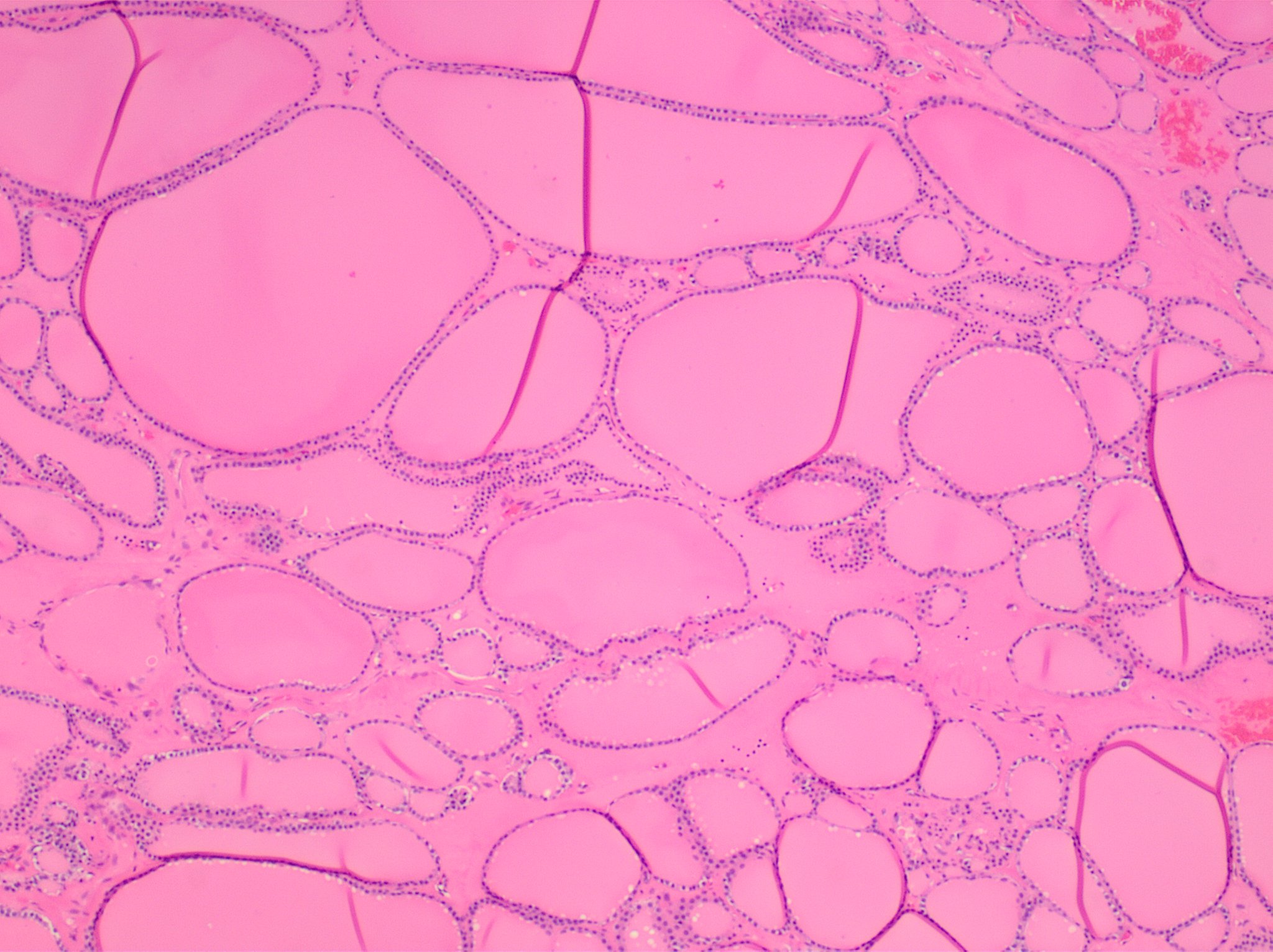 Diffuse thyroid hyperplasia typically shows variably sized follicles.
 -
 Thyroid hyperplasia with a hyperplastic nodule. It characteristically has no capsule (distinguishing it from thyroid adenoma or carcinoma).
 -
 The follicular linings may be thickened, with papillary projections (but lack nuclear features of papillary thyroid carcinoma).
 -
 The epithelium may also be flattened by enlarged follicles.
 -
 It can show hypercellular or microfollicular areas. There is no surrounding capsule (in contrast, a thyroid follicular adenoma or thyroid follicular carcinoma is generally encapsulated).
 -
 As shown, it can have mildly enlarged nuclei with mildly clumped chromatin, and clear cytoplasms, but cellular characteristics of papillary thyroid carcinoma are absent.
 -
 Thyroid hyperplasia with a Sanderson polster, which is a group of small follicles that protrude into the lumen of a larger follicle. It should not be confused for papillary projections.
 -
 Various forms of degeneration are typically seen, with various forms pictured.
Reporting
Example:
Right and left thyroid lobe, total thyroidectomy:
|
Notes
- ↑ For a full list of contributors, see article history. Creators of images are attributed at the image description pages, seen by clicking on the images. See Patholines:Authorship for details.
Main page
References
Image sources