Peripheral blood smear
Author:
Mikael Häggström [note 1]
Look at and comment separately on white blood cells, red blood cells and platelets.
Red blood cells
| RDW in adults[1] | Terminology[1] |
|---|---|
| 14.5% - 18% | Mild anisocytosis |
| 18% - 26% | Moderate anisocytosis |
| > 26% | Severe anisocytosis |
When available automatic quantification of mean corpuscular volume (MCV) and red blood cell distribution width (RDW), usually as part of CBC panel, generally decides whether you will call the sample "normocytic" versus "microcytic"/"macrocytic" and/or "anisocytotic", even if it is not clearly visible in the microscope. If automated values are not available, compare RBC sizes to lymphocyte nuclei, which should normally be the same size.
Look for poikilocytosis (red blood cells of abnormal shapes):
-
 Acanthocytes ("spur cells")
 -
 Echinocytes ("burr cells"), distinguished from acanthocytes by having more equally distributed and rounded projections. They do not need mention in the report.
 -
 Elliptocytes


Schistocytes (fragmented parts red blood cells) only needs mentioning if constituting over 1% of red blood cells or at least 2 per oil immersion field.
-
 When seeing what appears to a platelet overlying a red blood cells (as pictured), confirm that there is a halo.
 -
 Otherwise, make sure it is not malaria (appearance at different intraerythrocytic blood stages is pictured)...
 -
 ...or babesia.


Platelets
If CBC is performed, use count to determine whether platelets are "normal in number" or whether there is "thrombocytopenia" or "thrombocytosis". If no CBC, count platelets within a high power oil immersed field, which should normally be 8 to 20.
In thrombocytopenia from automatic counting, look in particular for clumping of platelets (which can cause a falsely low automatic platelet count). If present, check with the lab if it was sent in EDTA (which may cause artefactual clumping) and ask to have a blood sample sent in sodium citrate instead. Also, look for satellitosis (platelets attached around white blood cells).
White blood cells
Look for:
-
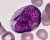 Blast cells, generally having large nucleus, immature chromatin, a prominent nucleolus, scant cytoplasm and few or no cytoplasmic granules. This example has an Auer rod (to the left of the nucleus).
 -
![Hypersegmented neutrophil. This is abnormal when more than half of neutrophils have at least 4 segments, or more than 5% of neutrophils have more than 5 segments.[2]](https://upload.wikimedia.org/wikipedia/commons/thumb/e/ea/Hypersegmented_neutrophil.jpg/250px-Hypersegmented_neutrophil.jpg) Hypersegmented neutrophil. This is abnormal when more than half of neutrophils have at least 4 segments, or more than 5% of neutrophils have more than 5 segments.[2]
Hypersegmented neutrophil. This is abnormal when more than half of neutrophils have at least 4 segments, or more than 5% of neutrophils have more than 5 segments.[2]

![Hypersegmented neutrophil. This is abnormal when more than half of neutrophils have at least 4 segments, or more than 5% of neutrophils have more than 5 segments.[2]](/File:Hypersegmented_neutrophil.jpg)
Notes
- ↑ For a full list of contributors, see article history. Creators of images are attributed at the image description pages, seen by clicking on the images. See Patholines:Authorship for details.
Main page
References
Image sources