Kidney autopsy
Author:
Mikael Häggström [note 1]
Contents
Comprehensiveness
On this resource, the following formatting is used for comprehensiveness:
- Minimal depth
- (Moderate depth)
- ((Comprehensive))
Autopsy cutting

In non-forensic autopsy, on each side:
- After evaluating the adrenal gland, dissect the renal fascia and perirenal fat laterally, and make an incision in the renal capsule. The renal capsule can then generally be loosened by hand. Note the surface texture. (Determine the color and consistency of the kidney.)
- Dissect the kidney in the coronal plane, towards the hilum. Inspect the cut surfaces.
Gross report
The kidneys are equally sized / (of normal size, with a total weight of ___ g)((a weight of ___ g on the right side and ___ g on the right)).
| Sex | Weight, reference range[note 2] | ||
| Right kidney | Left kidney | Total | |
| Men[2] | 80–160 g (2.8–5.6 oz) | 80–175 g (2.8–6.2 oz) | 160-335g (5.6-12.8 oz) |
| Women[3] | 40–175 g (1.4–6.2 oz) | 35–190 g (1.2–6.7 oz) | 75-365g (2.6-12.9 oz) |
(No abnormal adhesions between the kidneys and surrounding fibrous capsules.)
The kidneys have smooth surfaces/ {{<<Finely / Coarsely>> granular brown surface, possibly indicating benign nephrosclerosis. There are a few cysts on the surface containing clear fluid}}. Cut surfaces have well-defined medulla, cortex, and papillae. {{The cortices and/or medullas are narrowed and congested. The papillary portions are intact.}}
The renal pelvis and ureters are unremarkable /( Renal pelvis and ureters have normal calibers, with non-irritated mucosal surfaces and open lumens).
Fixation
Generally 10% neutral buffered formalin.
See also: General notes on fixation
Microscopic evaluation

Findings
The main findings to look for:[5]
Glomerular findings
- Diffuse and/or nodular mesangial deposits:
Nodular glomerulosclerosis, prompting a consideration of diabetic nephropathy, see section below.[5]
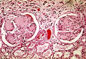
A glomerulus with total hyalinization (also called a globally sclerotic glomerulus). People aged ≤40 should have less than 1% - 10% globally sclerotic glomeruli, and less than 30% by age 80.[6] An increased amount indicates mainly hypertensive nephropathy, see section below.[6]
- Mesangial hypercellularity (which has been defined as more than 3 cell nuclei per peripheral mesangial area on a 4-μm thick tissue section), prompting a consideration of diabetic nephropathy, see section below.[5]
- Endocapillary hypercellularity, prompting a consideration of diabetic nephropathy, see section below.[5]
- Glomerular basement membrane thickening and/or duplication, prompting a consideration of diabetic nephropathy, see section below.[5]
Proteinaceous material in Bowman's space, see Proteinaceous material in Bowman's space
- Thrombi
Tubulointerstitial findings
- Interstitial inflammation
- Cytologic atypia in the tubular epithelial cells
- Crystals
- Atypical or pigmented casts
Vascular findings
- Thrombi
- Mural deposition of amorphous material
- Vasculitis
- Congestion
Intimal thickening with an onion skin-like appearance, indicating mainly hypertensive nephropathy, see section below.
Histopathology of arteriolar hyalinosis, being a component of nephroarteriolosclerosis, also indicating mainly hypertensive nephropathy, see section below.
Diagnoses
If indicated by findings above:
Diabetic nephropathy
For alterations in glomerular matrix and/or cellularity, the most common cause is diabetic nephropathy, and typically presents as glomerular enlargement, mesangial sclerosis, basement membrane thickening and and arteriolar hyalinosis.[5]
Hypertensive nephropathy
Hypertensive nephropathy has two types:
- Benign hypertensive nephrosclerosis: Characterized by arterial or arteriolar hyalinosis, intimal fibrosis, or medial hypertrophy.[7]
- Malignant hypertensive nephrosclerosis: Characterized by fibrinoid necrosis (acute stage) or myointimal cell proliferation, usually with an “onion-skinning” appearance (chronic stage).[7]
Report
- Findings and if they are consistent with already known diagnoses.
Further information: Autopsy
Notes
- ↑ For a full list of contributors, see article history. Creators of images are attributed at the image description pages, seen by clicking on the images. See Patholines:Authorship for details.
- ↑ Renal weight range is the standard reference range, that is, defined as the interval between which 95% of values of a reference population fall into.
Main page
References
- ↑ Guenevere Rae, Ph.D., William Newman, M.D., Supriya Donthamsetty, M.D., Robin McGoey, M.D.. The Cadaver’s Kidney P.G.. Retrieved on 2021-05-18.
- ↑ Standard reference range: Molina, D. Kimberley; DiMaio, Vincent J.M. (2012). "Normal Organ Weights in Men ". The American Journal of Forensic Medicine and Pathology 33 (4): 368–372. doi:. ISSN 0195-7910.
- ↑ Standard reference range: Molina, D. Kimberley; DiMaio, Vincent J. M. (2015). "Normal Organ Weights in Women ". The American Journal of Forensic Medicine and Pathology 36 (3): 182–187. doi:. ISSN 0195-7910.
- ↑ Mandolin S. Ziadie, M.D.. Simple cysts. Pathology Outlines. Topic Completed: 1 November 2011. Minor changes: 1 October 2019
- ↑ 5.0 5.1 5.2 5.3 5.4 5.5 Perrone, Marie E; Chang, Anthony; Henriksen, Kammi J (2017). "Medical renal diseases are frequent but often unrecognized in adult autopsies ". Modern Pathology 31 (2): 365–373. doi:. ISSN 0893-3952.
- ↑ 6.0 6.1 Jean L. Olson. Renal Disease Caused by Hypertension. Abdominal Key. Citing:
- Kaplan C, Pasternack B, Shah H, Gallo G (1975). "Age-related incidence of sclerotic glomeruli in human kidneys. ". Am J Pathol 80 (2): 227-34. PMID 51591. PMC: 1912918. Archived from the original. .
- Kappel, Birgitte; Olsen, Steen (1980). "Cortical interstitial tissue and sclerosed glomeruli in the normal human kidney, related to age and sex ". Virchows Archiv A Pathological Anatomy and Histology 387 (3): 271–277. doi:. ISSN 0340-1227. - ↑ 7.0 7.1 Liang, Shaoshan; Le, Weibo; Liang, Dandan; Chen, Hao; Xu, Feng; Chen, Huiping; Liu, Zhihong; Zeng, Caihong (2016). "Clinico-pathological characteristics and outcomes of patients with biopsy-proven hypertensive nephrosclerosis: a retrospective cohort study ". BMC Nephrology 17 (1). doi:. ISSN 1471-2369.
Image sources


