
Liver
Author:
Mikael Häggström [note 1]
Comprehensiveness
On this resource, the following formatting is used for comprehensiveness:
- Minimal depth
- (Moderate depth)
- ((Comprehensive))
Tissue sampling
- Autopsy
- Liver biopsy
Fixation
Generally 10% neutral buffered formalin. Non–formalin-fixed tissue may be needed for tests such as microbiological analysis or copper quantification studies.[1]
Gross processing in autopsy
- ((Measure the distance from the liver edge to the right costal margin.))
- Inspect the color and texture of the surfaces, including external and cut surfaces. Potential pathologies:
-
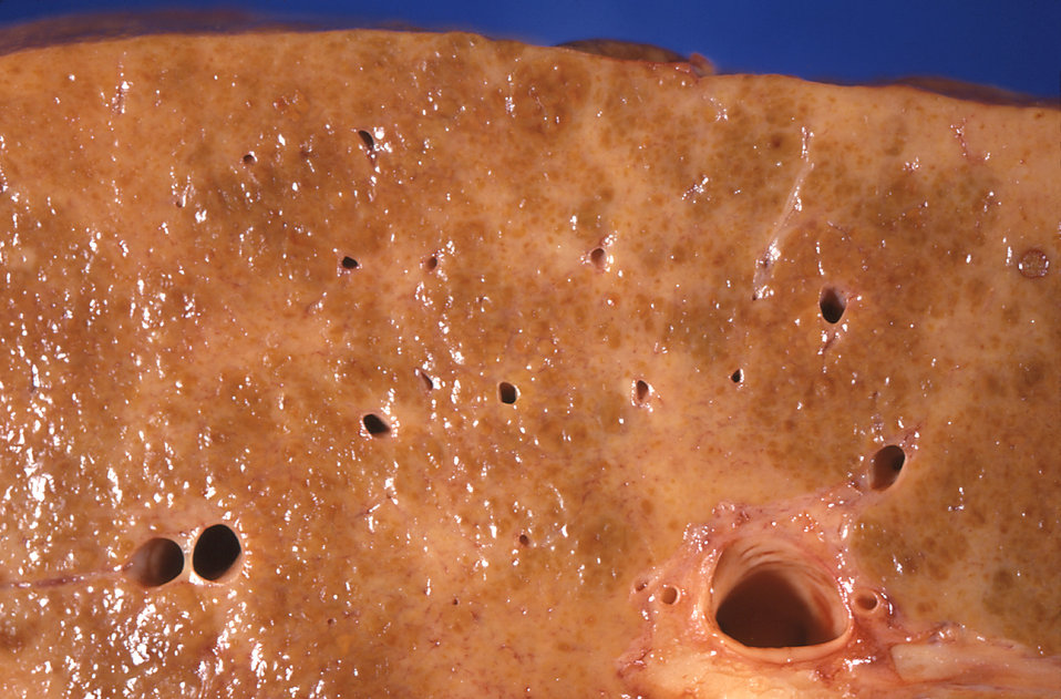 Diffuse areas of pallor in cirrhosis, see Cirrhosis
 -
.jpg) Pale macronodules of cirrhosis, see Cirrhosis
Pale macronodules of cirrhosis, see Cirrhosis -
.jpg) Nutmeg texture of congestive hepatopathy
Nutmeg texture of congestive hepatopathy -
.jpg) Liver metastases
Liver metastases
- Look for any focal change in the liver volume, mainly any tumor. If found: Further information: Liver tumor
- Determine liver weight. The standard reference range for men is 970–1,860 g (2.14–4.10 lb)[2] and for women 600–1,770 g (1.32–3.90 lb).[3]
- Make consecutive liver slices, such as in the sagittal or coronal plane.
Gross report in autopsy
- ((Distance from the liver edge to the right costal margin.))
- Weight. If abnormally low or high, preferably include the reference range.
- Color and texture of cut surfaces
- Any focal change
Example:
| Minimal | More comprehensive | Normal ranges |
| The liver weighs ___g. | The liver is << of normal size / {{enlarged}}, at ___g. | [[Men: 970-1860 g.[2] Women 600-1770g.[3].]] |
The liver surface is <<smooth ((and glistening)) {{/deformed by small and large nodules}}((, and is <<light tan / dark brown>> in color)). << Normal/ {{/ firm}} consistency. Cut surface[note 2] is normal / << (shows normal homogeneous brown parenchyma) / {{Yellowish color, indicating steatosis}} / {{dark nutmeg similar paths, indicating congestion}}. (No focal changes.)((The liver edge is _cm below the right costal margin.))
Microscopic evaluation

A general screening includes (with further information in sections below):
- Looking at the referral/requisition form (and looking at the medical records) for particular conditions to look for or evaluate. (Perform a severity grading of previously known liver diseases.)
- Commonly, this includes to quantify any cirrhosis, at least if the patient had alcohol abuse.
- Steatosis is also common.
- Signs of acute liver failure.
- Signs of malignancy. If a tumor is found: Further information: Liver tumor
- Signs of congestive hepatopathy (indicating heart failure).
- Signs of inflammation at least around the portal triads.
Cirrhosis
-
![Steatohepatitis with mild fibrosis (van Gieson's stain).[1]](https://upload.wikimedia.org/wikipedia/commons/a/af/Histopathology_of_steatohepatitis_with_mild_fibrosis_in_the_form_of_fibrous_expansion_(van_Gieson).jpg) Steatohepatitis with mild fibrosis (van Gieson's stain).[1]
Steatohepatitis with mild fibrosis (van Gieson's stain).[1] -
![Histopathology of steatohepatitis with moderate fibrosis (van Gieson's stain).[1]](https://upload.wikimedia.org/wikipedia/commons/d/da/Histopathology_of_steatohepatitis_with_moderate_fibrosis,_with_thin_fibrous_bridges_(van_Gieson).jpg) Histopathology of steatohepatitis with moderate fibrosis (van Gieson's stain).[1]
Histopathology of steatohepatitis with moderate fibrosis (van Gieson's stain).[1]
Further information: Cirrhosis
Steatosis
At least classify steatosis by severity:

-
 Note the presence of microvesicular steatosis, as it is usually more harmful. It shows foamy hepatocytes (two annotated by arrows), as opposed to the more common macrovesicular steatosis (insert).
 -
 Note any particular pattern of the steatosis, mainly centrilobular (pictured) or periportal.
Signs of acute liver failure
-
![Lobular disarray and associated lymphocytic inflammation, acidophil acidophil body formation (arrow) and bilirubinostasis, suggesting acute hepatitis.[1]](https://upload.wikimedia.org/wikipedia/commons/e/e6/Histopathology_of_acute_hepatitis.jpg) Lobular disarray and associated lymphocytic inflammation, acidophil acidophil body formation (arrow) and bilirubinostasis, suggesting acute hepatitis.[1]
Lobular disarray and associated lymphocytic inflammation, acidophil acidophil body formation (arrow) and bilirubinostasis, suggesting acute hepatitis.[1] -
![Shock liver, showing its hallmark[4] pathologic finding centrilobular necrosis but viable periportal hepatocytes. The necrotic hepatocytes are seen as slightly more eosinophilic (red) and discohesive.](https://upload.wikimedia.org/wikipedia/commons/a/ac/Histopathology_of_shock_liver_(intermediate_magnification),_annotated.jpg) Shock liver, showing its hallmark[4] pathologic finding centrilobular necrosis but viable periportal hepatocytes. The necrotic hepatocytes are seen as slightly more eosinophilic (red) and discohesive.
Shock liver, showing its hallmark[4] pathologic finding centrilobular necrosis but viable periportal hepatocytes. The necrotic hepatocytes are seen as slightly more eosinophilic (red) and discohesive. -
![Massive hepatic necrosis: Liver cell dropout, residual hepatocytes and intact portal tract pattern.[5]](https://upload.wikimedia.org/wikipedia/commons/6/63/Histopathology_of_massive_hepatic_necrosis_in_hepatitis_B.jpg) Massive hepatic necrosis: Liver cell dropout, residual hepatocytes and intact portal tract pattern.[5]
Massive hepatic necrosis: Liver cell dropout, residual hepatocytes and intact portal tract pattern.[5]
Congestive hepatopathy

- Acute hepatic congestion shows dilated sinusoidal capillaries predominantly in zone 3 of the hepatic acinus.[7]
- Typical findings of chronic hepatic congestion are atrophy of hepatocytes in zone 3, perisinusoidal edema, thrombosis and hemorrhage. Chronic congestion typically displays perivenous and perisinusoidal fibrosis, with fibrous septa that bridge central hepatic veins. In contrast, other causes of distortion and cirrhosis typically have fibrous septa predominantly between portal triads. However, nonalcoholic steatohepatitis also may show perisinusoidal fibrosis in early stages; in later stages, the fibrosis tends to be in the portal triad. Cirrhosis develops in the final stages of congestive hepatopathy. Regenerating hepatocytes tend to grow in a sleevelike pattern along portal tracts, resulting in a nodular liver with preserved portal triads and obliterated or fibrosed hepatic veins, a pattern called "reverse lobulation". This pattern can also be seen in venous obstruction due to Budd-Chiari syndrome.[8]
Hemosiderin
-
 Look for Kupffer cells with significant hemosiderin deposition (shown next to a hepatocyte with lipofuscin pigment.
 -
 Iron stain can highlight the hemosiderin (blue stain) in unclear cases.
 -
![Hepatocyte lipofuscin alone is of no real pathologic importance and does not warrant a mention in the report.[9]](https://upload.wikimedia.org/wikipedia/commons/2/28/Histology_of_hepatocyte_lipofuscin.jpg) Hepatocyte lipofuscin alone is of no real pathologic importance and does not warrant a mention in the report.[9]
Hepatocyte lipofuscin alone is of no real pathologic importance and does not warrant a mention in the report.[9]
Report
- Presence of any liver disease
- (Quantification of its severity.)
- ((Even absence of hepatitis, malignancy, congestive hepatopathy and/or steatosis.))
Notes
- ↑ For a full list of contributors, see article history. Creators of images are attributed at the image description pages, seen by clicking on the images. See Patholines:Authorship for details.
- ↑ "Cut surface shows..." may alternatively be expressed as "On sectioning, the parenchyma is..."
Main page
References
- ↑ 1.0 1.1 1.2 1.3 Boyd, Alexander; Cain, Owen; Chauhan, Abhishek; Webb, Gwilym James (2020). "Medical liver biopsy: background, indications, procedure and histopathology
". Frontline Gastroenterology 11 (1): 40–47. doi:. ISSN 2041-4137.
- "This is an open access article distributed in accordance with the Creative Commons Attribution 4.0 Unported (CC BY 4.0) license"
- ↑ 2.0 2.1 Standard reference range: Molina, D. Kimberley; DiMaio, Vincent J.M. (2012). "Normal Organ Weights in Men ". The American Journal of Forensic Medicine and Pathology 33 (4): 368–372. doi:. ISSN 0195-7910.
- ↑ 3.0 3.1 Standard reference range: Molina, D. Kimberley; DiMaio, Vincent J. M. (2015). "Normal Organ Weights in Women ". The American Journal of Forensic Medicine and Pathology 36 (3): 182–187. doi:. ISSN 0195-7910.
- ↑ Ciobanu AO, Gherasim L (2018). "Ischemic Hepatitis - Intercorrelated Pathology. ". Maedica (Bucur) 13 (1): 5-11. PMID 29868133. PMC: 5972787. Archived from the original. .
- ↑ Xue, Ran; Zhu, Yueke; Liu, Hui; Meng, Qinghua (2019). "The clinical parameters for the diagnosis of hepatitis B virus related acute-on-chronic liver failure with sepsis
". Scientific Reports 9 (1). doi:. ISSN 2045-2322.
-"This article is licensed under a Creative Commons Attribution 4.0 International License" - ↑ Shah, Shailja C.; Sass, David A. (2015). "“Cardiac Hepatopathy”: A Review of Liver Dysfunction in Heart Failure
". Liver Research - Open Journal 1 (1): 1–10. doi:. ISSN 23794038.
-"This is an open access article distributed under the Creative Commons Attribution 4.0 International License (CC BY 4.0)," - ↑ . Acute Hepatic Congestion. Pathway Medicine. Retrieved on 2020-03-06.
- ↑ Wells, Michael L.; Fenstad, Eric R.; Poterucha, Joseph T.; Hough, David M.; Young, Phillip M.; Araoz, Philip A.; Ehman, Richard L.; Venkatesh, Sudhakar K. (2016). "Imaging Findings of Congestive Hepatopathy ". RadioGraphics 36 (4): 1024–1037. doi:. ISSN 0271-5333.
- ↑ . The Internet Pathology Laboratory for Medical Education. The University of Utah Eccles Health Sciences Library. Retrieved on 2020-12-18.
Image sources