Lung tumor
Author:
Mikael Häggström [note 1]
Contents
Comprehensiveness
On this resource, the following formatting is used for comprehensiveness:
- Minimal depth
- (Moderate depth)
- ((Comprehensive))
Presentations
- Bronchial lavage
- Lung needle biopsy
- Lung wedge resection or lobectomy
- Lung autopsy
Gross processing
As per presentation above.
Microscopic evaluation
Medical imaging provides a major clue as to whether a lung tumor is benign or malignant, where lesions smaller than 2 cm are likely to be benign, whereas lesions larger than 2 cm are malignant (that is, lung cancer) in 85% of cases.[1]
Benign tumors
Subsequently distribution of benign tumors and lung cancers, respectively, are as follows:[1]
Benign lung tumors:
- Hamartomas - 76%
- Benign fibrous mesothelioma/solitary fibrous tumor (SFT) - 12.3%
- Inflammatory pseudotumor (IPT) - 5.4%
- Lipoma - 1.5%
- Leiomyoma - 1.5%
- Other - 3.3%
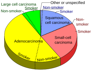
Lung cancers
Lung adenocarcinoma, with lepidic pattern shown, wherein tumors cells cover alveolar walls.
Lung adenocarcinoma, with solid pattern.
Squamous-cell carcinoma (SCC) of the lung. Typical squamous-cell carcinoma cells are large with abundant eosinophilic cytoplasm and large, often vesicular, nuclei.[3]
Small-cell carcinoma, with typical findings.[4]

Whereas large cell carcinoma is more often histologically distinct, adenocarcinoma and SCC may look alike. In such cases, an immunohistochemistry panel of TTF1, CK5/6, and p63 can be used to distinguish the two.[6][7]
Further workup
For primary lung non-small cell carcinoma (NSCLC) stages IB - IV (such as being more than 3 cm in size), generally perform full next generation sequencing panel (DNA and RNA) with PDL-1 immunostaining. For an advanced stage NSCLC that is not a candidate for biopsy or re-biopsy, a viable alternative is “liquid biopsy” on peripheral blood for circulating tumor DNA.[8]
Notes
- ↑ For a full list of contributors, see article history. Creators of images are attributed at the image description pages, seen by clicking on the images. See Patholines:Authorship for details.
Main page
References
- ↑ 1.0 1.1 Alain C. Borczuk (2008). "Benign Tumors and Tumorlike Conditions of the Lung ". Archives of Pathology & Laboratory Medicine 132 (7). Archived from the original. .
- ↑ Kuroki, Masaomi; Nakata, Hiroshi; Masuda, Toshifumi; Hashiguchi, Norihisa; Tamura, Shozo; Nabeshima, Kazuki; Matsuzaki, Yasunori; Onitsuka, Toshio (2002). "Minute Pulmonary Meningothelial-like Nodules: High-Resolution Computed Tomography and Pathologic Correlations ". Journal of Thoracic Imaging 17 (3): 227–229. doi:. ISSN 0883-5993.
- ↑ Dr Nicholas Turnbull, A/Prof Patrick Emanual (2014-05-03). Squamous cell carcinoma pathology. DermNetz.
- ↑ Image by Mikael Häggström, MD. Source for findings: Caroline I.M. Underwood, M.D., Carolyn Glass, M.D., Ph.D.. Lung - Small cell carcinoma. Pathology Outlines. Last author update: 20 September 2022}}
- ↑ Image by Mikael Häggström, MD. Source for significance: Bejarano PA, Mousavi F (2003). "Incidence and significance of cytoplasmic thyroid transcription factor-1 immunoreactivity. ". Arch Pathol Lab Med 127 (2): 193-5. doi:. PMID 12562233. Archived from the original. .
- ↑ Inamura K (2018). "Update on Immunohistochemistry for the Diagnosis of Lung Cancer. ". Cancers (Basel) 10 (3). doi:. PMID 29538329. PMC: 5876647. Archived from the original. .
- ↑ Affandi KA, Tizen NMS, Mustangin M, Zin RRMRM (2018). "p40 Immunohistochemistry Is an Excellent Marker in Primary Lung Squamous Cell Carcinoma. ". J Pathol Transl Med 52 (5): 283-289. doi:. PMID 30235512. PMC: 6166010. Archived from the original. .
- ↑ . National Comprehensive Cancer Network (NCCN) Clinical Practice Guidelines in Oncology (NCCN Guidelines) - Non-Small Cell Lung Cancer. Version 3.2024. Section: Principles of molecular and biomarker analysis (2024-03-12).
Image sources
- ↑ Image(s) by: Mikael Häggström, M.D. Public Domain
- Author info
- Reusing images


