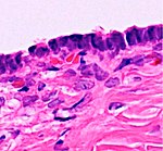
Ovarian cyst
Jump to navigation
Jump to search
Author:
Mikael Häggström [note 1]
Contents
Presentations
Typically a cyst with more or less of the ovary.
Comprehensiveness
On this resource, the following formatting is used for comprehensiveness:
- Minimal depth
- (Moderate depth)
- ((Comprehensive))
- Other legend
<< Decision needed between alternatives separated by / signs >>
{{Common findings / In case of findings}}
[[Comments]]
Link to another page
Gross processing
The specimen is generally not inked.
Gross report
Example:
| (Labeled - ___.) The specimen is received in formalin and consists of an ovary with a cyst, and attached portion of the fallopian tube, in total measuring ___. The capsule of the ovary and the cyst is tan-pink and smooth. The cyst is << intact / collapsed>>{{, with a wall defect measuring _ cm}}. Upon {{further}} opening the contents of the cyst consists of ___. The cyst lining is tan-pink and smooth, with a wall thickness of __. There are no papillary excrescences. Cut sections of the ovary reveal solid, white and whorled parenchyma, and no gross lesions. The attached fallopian tube fragment measures ___, and displays fimbria. Cut sections of the tube reveal a patent lumen. Representative sections are submitted for microscopic examination in __ cassettes. |
Microscopic evaluation
If there are solid components, evaluate those as ovarian tumors.
Attempt to classify cysts into either of the following:
| Type | Subtype | Typical microscopy findings | Image |
|---|---|---|---|
| Functional cyst | Follicular cyst |
|

|
| Corpus luteum cyst | 
| ||
| Cystadenoma | Serous cystadenoma | Cyst lining consisting of a simple epithelium, whose cells may be either:[3]
|

|
| Mucinous cystadenoma | Lined by a mucinous epithelium | 
| |
| Mature cystic teratoma (dermoid) |
Well-differentiated components from at least two germ layers (ectoderm, mesoderm and/or endoderm).[4] | 
| |
| Endometriosis | At least two of the following three criteria:[5]
|

| |
| Borderline tumor | Atypical epithelial proliferation without stromal invasion.[6] | 
| |
| Ovarian cancer | Many different types, but generally severe dysplasia/atypia and invasion. | ||
| Simple cyst / simple squamous cyst |
Simple squamous epithelium and not conforming to diagnoses above | 
| |
Notes
- ↑ For a full list of contributors, see article history. Creators of images are attributed at the image description pages, seen by clicking on the images. See Patholines:Authorship for details.
Main page
References
- ↑ 1.0 1.1 Mohiedean Ghofrani. Ovary - nontumor - Nonneoplastic cysts / other - Follicular cysts. Pathology Outlines. Topic Completed: 1 August 2011. Revised: 5 March 2020
- ↑ 2.0 2.1 2.2 Aurelia Busca, Carlos Parra-Herran. Ovary - nontumor - Nonneoplastic cysts / other - Corpus luteum cyst (CLC). Pathology Outlines. Topic Completed: 1 November 2016. Revised: 5 March 2020
- ↑ Shahrzad Ehdaivand, M.D.. Ovary tumor - serous tumors - Serous cystadenoma / adenofibroma / surface papilloma. Pathology Outlines. Topic Completed: 1 June 2012. Revised: 5 March 2020
- ↑ Sahin, Hilal; Abdullazade, Samir; Sanci, Muzaffer (2017). "Mature cystic teratoma of the ovary: a cutting edge overview on imaging features ". Insights into Imaging 8 (2): 227–241. doi:. ISSN 1869-4101.
- ↑ Aurelia Busca, Carlos Parra-Herran. Ovary - nontumor - Nonneoplastic cysts / other - Endometriosis. Pathology Outlines. Topic Completed: 1 August 2017. Revised: 5 March 2020
- ↑ Lee-may Chen, MDJonathan S Berek, MD, MMS. Borderline ovarian tumors. UpToDate. This topic last updated: Feb 08, 2019.
Image sources

